Investigations

Sound Waves and Heart Health: The Echocardiogram Explained

An echocardiogram, often referred to as an “echo,” is a fundamental diagnostic tool in the field of cardiology. This non-invasive procedure harnesses the power of sound waves to provide a comprehensive assessment of the heart’s structure and function. Widely utilized by healthcare professionals, the echocardiogram is an invaluable resource for diagnosing and monitoring a spectrum of cardiac conditions. This article delves into echocardiography, shedding light on its significance in heart health.


How is an Echocardiogram Performed?

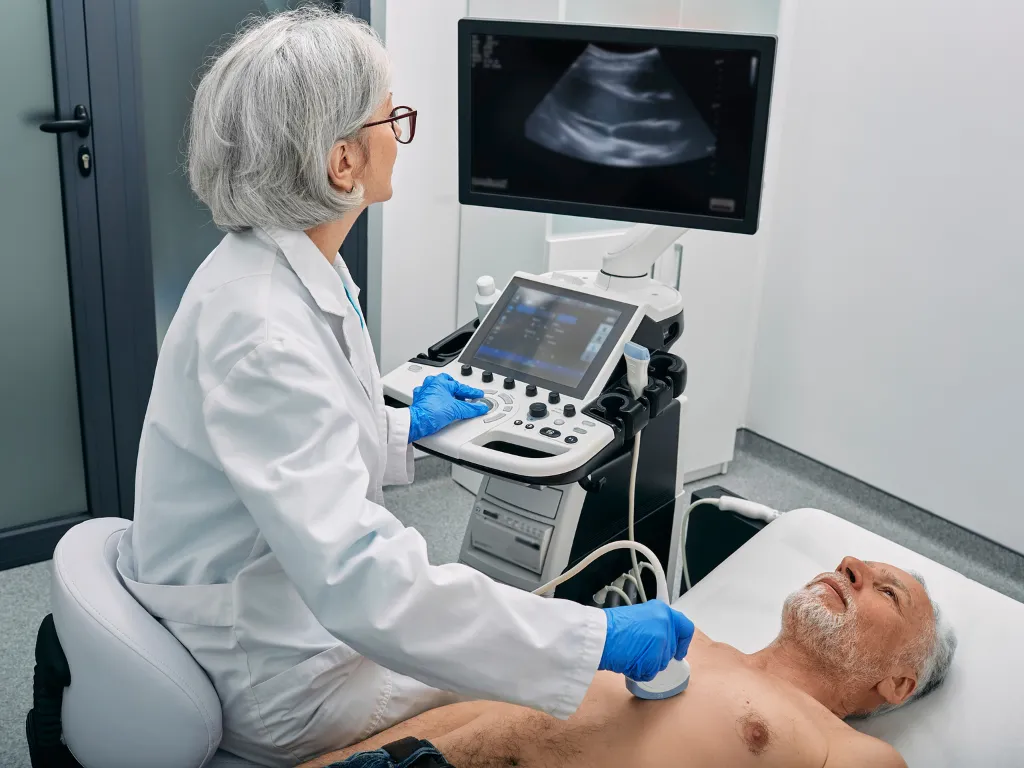
The procedure involves using a small transducer, which resembles a handheld device and is placed on the chest. The sonographer performing the test carefully positions the transducer so that it is directed toward the heart. Several different views are acquired from different positions around the chest wall.

Sound Waves and Heart Health: The Echocardiogram Explained Heart Matters

Sound Waves Unveiling the Heart’s Secrets

The magic behind an echocardiogram lies in the high-frequency sound waves emitted by the transducer. These sound waves, which are beyond the range of human hearing, serve as the silent messengers of cardiac health. When the transducer releases these sound waves, they travel through the patient’s chest and ultimately reach the heart.

Echoes Paint a Picture

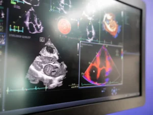
These sound waves bounce back or “echo” to the transducer as they contact the heart’s structures. This is where the true marvel of echocardiography unfolds. The transducer skillfully captures the returning echoes and transforms them into real-time images displayed on a monitor.

Revealing the Heart’s Inner Workings

The images generated during an echocardiogram give clinicians an unparalleled view of the heart’s inner workings. These images vividly showcase the heart’s chambers, valves, and the dynamic flow of blood through the organ. This comprehensive visual data is invaluable in assessing the heart’s overall health, enabling healthcare professionals to detect potential abnormalities or issues.
Sound Waves and Heart Health: The Echocardiogram Explained Heart Matters

There are several different applications of echocardiograms, each with its purpose and benefits. Some common types include:

  • Transthoracic Echocardiogram (TTE): Among the most commonly performed echocardiograms, TTE is conducted by placing the transducer directly on the skin over the chest. This approach offers a comprehensive view of the heart’s structure and function. TTE is a versatile and widely utilized diagnostic tool, providing valuable insights into cardiac health.
  • Stress Echocardiogram: Combining the power of an echocardiogram with either exercise or medication that induces stress on the heart, this test allows healthcare professionals to assess how the heart responds under duress. Stress echocardiograms are invaluable for evaluating the heart’s performance during increased demand, diagnosing conditions such as coronary artery disease, or assessing the effectiveness of treatment plans.
  • Transesophageal Echocardiogram (TEE): This specialized echocardiogram involves the insertion of a slender tube equipped with a transducer down the throat and into the esophagus, which is positioned adjacent to the heart. This shares some principles with endoscopy, such as using a flexible tube inserted through the throat. However, it’s important to clarify that while TEE and endoscopy have similarities in their approach, their primary objectives and focus areas are distinctly different. The TEE aims to obtain a highly detailed image of the heart’s internal structures, particularly of the heart valves. TEE is typically employed when a clear and precise image is required or in cases where a standard Transthoracic Echocardiogram (TTE) may not provide the necessary level of detail.


    Risks of Echocardiography

    Echocardiography, a widely utilized diagnostic modality, relies on harmless ultrasound sound waves to provide detailed cardiac imaging without radiation exposure.

    However, the nature of potential risks associated with echocardiography varies based on the specific procedure:

    Transthoracic Echocardiogram (TTE): Typically, this procedure is painless and takes around 30 minutes to complete. Occasionally, when the sonographer requires firmer pressure to optimize image quality, individuals may experience momentary discomfort in the area of the chest where the probe is applied.

    Transesophageal Echocardiogram (TEE):  While transesophageal echocardiograms (TEEs) are generally considered safe and well-tolerated, it’s important to be aware of potential risks associated with this procedure. Patients may experience temporary throat soreness following the test, and in rare cases, the tube used during TEE may cause minor irritation to the throat’s interior. There is also a slight risk of difficulties in swallowing or temporary hoarseness. Additionally, some patients may encounter muscular spasms affecting the throat or lungs and minor bleeding in the throat region. Although uncommon, teeth, gums, or lips injuries have been reported. A rare yet serious complication involves esophageal perforation, characterized by the formation of a hole in the esophagus. Before undergoing a TEE, your doctor will take the time to discuss the potential risks and the benefits of the procedure, ensuring that you are well informed and comfortable with the decision.

    Sound Waves and Heart Health: The Echocardiogram Explained Heart Matters


    Conclusion

    In summary, echocardiography is a pivotal diagnostic modality in cardiology, providing essential insights into cardiac structure and function. Through procedures like Transthoracic Echocardiograms (TTE), Transesophageal Echocardiograms (TEE), and Stress Echocardiograms, healthcare professionals gain crucial data for diagnosing and managing cardiac conditions. Echocardiography’s non-invasive, radiation-free nature and real-time imaging capabilities underscore its importance in cardiac health. As cardiology advances, echocardiography plays a central role in improving patient outcomes and our understanding of heart health.

    Search articles

    Newsletter

    Subscribe for exclusive heart health resources, valuable tips, and the latest research updates.

    Editor's Picks

    Empowering heart health

    Subscribe to our newsletter and be the first to receive valuable insights, tips, and resources on heart health.

    other anti-anginals

    When first-line therapies for angina, such as beta blockers, calcium channel blockers, and nitrates, prove inadequate or are not well-tolerated, second-line therapies may be considered.
    Perhexiline is a unique medication that enhances the heart's ability to utilize fatty acids for energy, reducing its reliance on oxygen and lowering oxygen demand. This action helps improve blood flow and alleviates chest pain in some patients with refractory angina.
    Nicorandil is another second-line option with a dual mechanism of action. It opens potassium channels in smooth muscle cells, causing vasodilation and enhancing coronary blood flow. Additionally, nicorandil also stimulates nitric oxide release, further dilating blood vessels and reducing heart workload.
    Trimetazidine is an anti-ischemic agent that improves cardiac efficiency by enhancing glucose metabolism and shifting the heart's energy production to a more oxygen-efficient process. As second-line therapies, these medications offer alternative approaches for managing angina in individuals who do not respond adequately to first-line treatments or those experiencing side effects from other medications.

    lipid lowering therapies

    Lipid-lowering therapies play a critical role in managing coronary artery disease (CAD), a condition characterized by the narrowing of blood vessels that supply the heart. Among the most commonly discussed and debated classes of medications are statins, which effectively reduce cholesterol levels and are widely prescribed to lower the risk of cardiovascular events. Alongside statins, other medications like ezetimibe, fibrates, and niacin are also utilized to target specific aspects of lipid metabolism, such as cholesterol absorption, triglyceride levels, and raising high-density lipoprotein (HDL) cholesterol. Additionally, the introduction of medications that inhibit PCSK9, an enzyme involved in cholesterol metabolism, has provided a promising new approach to further lower LDL cholesterol levels. These PCSK9 inhibitors, such as Repatha (evolocumab), have shown significant efficacy in reducing LDL cholesterol levels in patients with CAD, especially for those who may not respond well to traditional therapies.

    Nitrates

    Nitrates are widely used to treat angina and provide quick relief for chest pain. Commonly available in the form of sublingual sprays or tablets, patches, and long-acting tablets, nitrates work by dilating blood vessels, allowing for increased blood flow and reduced resistance. This dilation eases the heart's workload, leading to a decreased demand for oxygen and prompt alleviation of angina symptoms. Sublingual nitrates act rapidly and are often used to provide immediate relief during angina attacks, while patches and long-acting tablets are employed for preventive purposes. However, nitrates may cause side effects such as headaches, dizziness, and flushing, which usually subside over time.

    calcium channel blockers

    Calcium channel blockers, including amlodipine, felodipine, cardizem (diltiazem), and verapamil, are commonly prescribed for the treatment of angina. These medications work by inhibiting the influx of calcium into the muscle cells of the heart and blood vessels, leading to their relaxation. As a result, blood vessels widen, promoting improved blood flow and reduced blood pressure. In the context of angina, this relaxation decreases the heart's workload, lowering the demand for oxygen and alleviating chest pain. Calcium channel blockers offer a valuable treatment option for individuals with angina, but it is essential to be aware of potential side effects, which may include headaches, dizziness, flushing, and ankle swelling.

    Beta blockers

    Beta blockers, such as metoprolol, propranolol, atenolol, carvedilol, and bisoprolol, play a crucial role in treating angina. By blocking certain receptors in the heart, they effectively reduce heart rate and the force of contraction, thereby easing the heart's workload. This mechanism of action leads to a decreased demand for oxygen, making beta blockers highly effective in relieving chest pain associated with angina. As with any medication, it's important to consider potential side effects, including tiredness, worsened asthma, erectile dysfunction in some males, and more vivid dreams during sleep. Consult your healthcare provider to determine the suitability of beta blockers for managing your angina and overall heart health.

    Anti-platelet Medications

    Anti-platelet medications play a crucial role in preventing blood clot formation, reducing the risk of serious cardiovascular events such as heart attacks and strokes. Among the widely used anti-platelet drugs are aspirin, clopidogrel, and ticagrelor.

    Aspirin: This well-known medication inhibits platelet activation, making it less likely for platelets to stick together and form clots. Aspirin is commonly used for primary and secondary prevention of heart attacks and strokes.

    Clopidogrel: As a potent anti-platelet agent, clopidogrel works by blocking specific receptors on platelets, preventing them from aggregating. It is often prescribed to patients with acute coronary syndrome, those undergoing stent procedures, and for some cases of peripheral arterial disease.

    Ticagrelor: Ticagrelor is another effective anti-platelet drug that works by inhibiting platelet activation. It is used in acute coronary syndrome, often given alongside aspirin to reduce the risk of heart-related events.