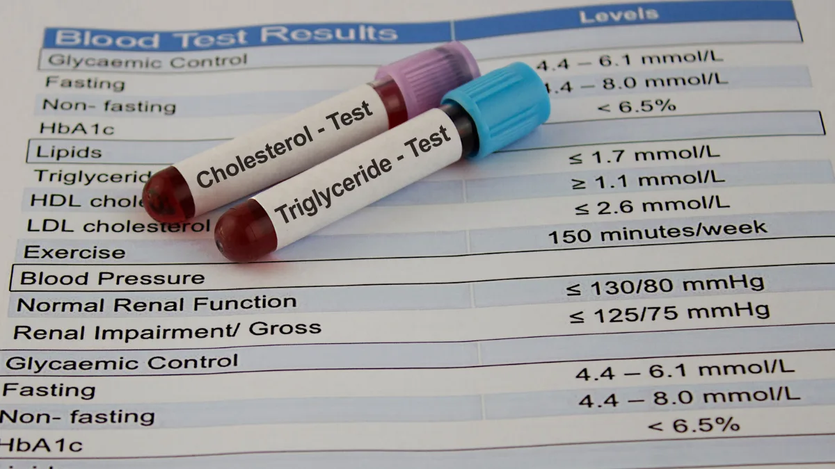



- Most heart-related blood tests do not require fasting. Fasting matters most for the fasting glucose and glucose tolerance tests, and is often recommended for cholesterol tests, particularly when triglycerides are measured.
- Fasting is often recommended before a cholesterol panel, because meals raise triglycerides and fasting keeps results consistent between tests. A normal meal changes LDL and HDL cholesterol only slightly, so a non-fasting sample is acceptable for many routine checks.
- The HbA1c test, which reflects average blood glucose over the previous two to three months, does not require fasting.
- When fasting is needed, it usually means no food or drinks other than water for 8 to 12 hours. Exact instructions vary between laboratories.
- Fasting can cause low blood sugar in people taking insulin or some diabetes tablets, which is why they are usually given specific instructions before a fasting test.
Few instructions cause as much confusion as “fast before your blood test.” Does black coffee count? What about morning tablets? And why does one cholesterol test come with fasting instructions while the next does not?
The short answer is that fasting matters a great deal for a few tests, matters in particular situations for cholesterol, and makes very little difference for most others. Knowing which is which can spare an uncomfortable morning and, for some people, avoid a genuine risk.
Why Food Affects Some Blood Tests
After a meal, sugars and fats from food pass into the bloodstream. For a few hours, the levels of some substances in the blood rise, while others barely change.
Glucose (blood sugar) and triglycerides are the two most affected. A test designed to measure a “resting” level of either can be misleading if taken soon after eating. Most other routine blood tests are affected so little by a normal meal that fasting makes no practical difference.
Which Heart-Related Tests Need Fasting?
The table below shows how fasting requirements typically apply to common blood tests in heart care. Individual laboratories may set their own requirements, and the instructions given with a test request take priority.
| Test | Fasting Usually Needed? | Why |
|---|---|---|
| Fasting glucose | Yes | Food raises blood glucose for several hours |
| Glucose tolerance test | Yes | Measures the response to a standard glucose drink from a fasting start |
| HbA1c | No | Reflects average glucose over months, not today’s meals |
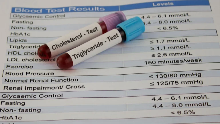
| Cholesterol panel, including triglycerides | Often recommended | Meals raise triglycerides, and fasting keeps results comparable from one test to the next |
| Iron studies | Sometimes | Some laboratories prefer a morning sample, as iron levels vary |
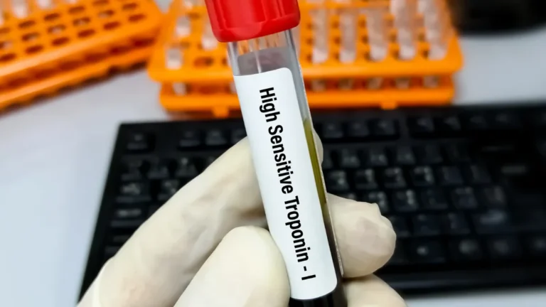
| Troponin and BNP | No | Often urgent, and not meaningfully affected by food |
| INR, kidney function, hs-CRP | Usually not | Routine checks are generally done without fasting |
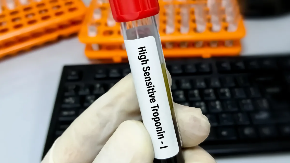
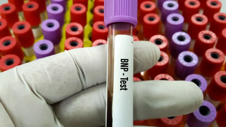
Tests used to assess chest pain or heart strain, such as troponin and BNP, are taken whenever they are needed, often in an emergency department. Fasting plays no part in them.
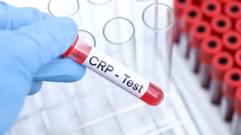
Routine monitoring tests, such as the INR used for people taking warfarin and the hs-CRP test, are generally done without fasting. For iron studies, used to check for iron deficiency, some laboratories prefer a morning sample because iron levels in the blood vary through the day.
Glucose Tests and Fasting
Blood glucose tests are where fasting matters most. They are used to check for diabetes and prediabetes, both important risk factors for heart disease.
Fasting glucose
A fasting glucose test measures blood sugar after a period without food. The American Diabetes Association defines fasting as no calorie intake for at least 8 hours. An overnight fast with a morning test is the usual arrangement.
Eating before this test can push the result into a range that looks like diabetes when it is not. That is why a fasting glucose result taken after breakfast cannot be interpreted in the usual way.
Glucose tolerance test
The oral glucose tolerance test shows how well the body clears sugar from the blood. After an overnight fast, a blood sample is taken, a standard drink containing 75 g of glucose is given, and a second sample is taken two hours later. The test takes a couple of hours in total and is commonly used in pregnancy.
HbA1c
The HbA1c test measures how much glucose has attached to red blood cells over the previous two to three months. Because it reflects a long-term average, a meal beforehand has no meaningful effect, and no fasting is needed.
HbA1c is reported in two ways around the world: as a percentage, or in millimoles per mole (mmol/mol). A result of 6.5% is the same as 48 mmol/mol.
Glucose results in both units
Glucose is reported in mmol/L in many countries and in mg/dL in others, including the United States. A result in mmol/L multiplied by 18 gives the value in mg/dL. The table shows the levels the American Diabetes Association uses to define diabetes.
| Test | Fasting? | Diabetes Range (mmol/L) | Diabetes Range (mg/dL) |
|---|---|---|---|
| Fasting glucose | Yes | 7.0 or higher | 126 or higher |
| Glucose tolerance test (2 hours) | Yes | 11.1 or higher | 200 or higher |
| Random glucose, with symptoms | No | 11.1 or higher | 200 or higher |
The range for prediabetes differs between organisations. The American Diabetes Association uses a fasting glucose of 5.6 to 6.9 mmol/L (100 to 125 mg/dL), while the World Health Organization uses 6.1 to 6.9 mmol/L (110 to 125 mg/dL). Unless a result is clearly abnormal, a diagnosis of diabetes is usually confirmed with a second test, and interpreting results is a matter for the requesting doctor.
Cholesterol Tests: When Fasting Helps
Cholesterol tests were traditionally done after fasting. A joint consensus statement from the European Atherosclerosis Society and the European Federation of Clinical Chemistry and Laboratory Medicine reviewed large studies comparing fasting and non-fasting results.
After normal meals, triglycerides rose by up to about 0.3 mmol/L (26 mg/dL), while total and LDL cholesterol fell by about 0.2 mmol/L (8 mg/dL). HDL cholesterol and lipoprotein(a) were not affected. Non-fasting results predicted heart disease risk just as well as fasting ones, so the statement recommends non-fasting samples for routine testing.
Why triglycerides are different
Triglycerides are the part of the cholesterol panel most affected by food. The average rise after a meal is small, but it can be much larger after a fatty meal, and in people with diabetes, excess abdominal weight, or already raised triglycerides.
This matters for LDL cholesterol too. In most laboratories, LDL is calculated from the other values, including triglycerides. The higher the triglyceride level, the less accurate that calculation becomes.
For these reasons, American guidelines state that a fasting sample is most helpful in several situations:
- When triglycerides have been high before, particularly above 4.5 mmol/L (400 mg/dL).
- When high triglycerides are being diagnosed or monitored.
- When there is a family history of early heart disease or an inherited cholesterol disorder.
- When checking how much risk remains for a person already on cholesterol-lowering treatment.
European guidance suggests a fasting repeat when a non-fasting triglyceride level is above 5 mmol/L (440 mg/dL). In everyday practice, many doctors request a fasting sample for cholesterol tests, particularly when triglycerides are part of the question. When a test request asks for fasting, that instruction is the one that applies.
I usually recommend fasting before a cholesterol test. I often request a full panel, including glucose and triglycerides, and fasting keeps results consistent, which makes monitoring over time more reliable.
Prof. Peter Barlis, Interventional Cardiologist
What Counts as Fasting?
Definitions vary. The American Diabetes Association uses at least 8 hours without calories, while European laboratory guidance recommends 12 hours. Water is permitted in both, and many laboratories encourage it, as good hydration can make blood easier to draw.
European laboratory guidance also advises against alcohol for 24 hours beforehand, and against coffee, tea, and smoking on the morning of the test. Chewing gum can affect some results too. Because practice differs between countries and laboratories, the instructions supplied with the test are the most reliable guide.
Medicines and Fasting
Advice about medicines on the morning of a fasting test varies. Most regular heart medicines are commonly taken as normal with water, but some laboratories and doctors ask for particular medicines to be delayed until after the sample. The requesting doctor or laboratory gives instructions for each person.
People with diabetes need particular care. Insulin and some diabetes tablets can cause low blood sugar when a meal is skipped, which can lead to shakiness, sweating, confusion, or fainting. For this reason, people taking these medicines are usually given specific instructions about timing their medicine and the test, and tests are often booked early in the morning.
Conclusion
Fasting matters for a small number of blood tests, chiefly the fasting glucose and glucose tolerance tests. For cholesterol tests, fasting is often recommended because it gives a clearer and more consistent triglyceride result, although a non-fasting sample is acceptable for many routine checks. For most other heart-related tests, including HbA1c, fasting makes little or no difference.
When fasting is required, the instructions that come with the test request are the best guide, and questions about medicines are worth raising with the requesting doctor or laboratory beforehand. A few minutes of clarification can prevent a wasted trip or an unreliable result.
Related Reading
- Understanding Your Cardiovascular Risk Factors
- Lipoprotein(a): The Inherited Heart Risk Most People Have Never Heard Of
- The Timing of Cholesterol-Lowering Medications: Does Nighttime Dosing Make a Difference?
- Heart Health in Asian Populations
- Dietary Fibre and Your Heart: Why This Nutrient Deserves More Attention