- Despite the word “block”, first-degree AV block is not a blockage. Every electrical signal still reaches the lower chambers of the heart. It simply takes slightly longer to get there.
- It is found on roughly 1 to 2% of ECGs in healthy adults, and is more common in athletes and with increasing age. In most people it causes no symptoms and is discovered by accident.
- Common causes include normal variation, high fitness levels, ageing of the conduction system, and several widely used heart medications.
- It generally needs no treatment. The situations that prompt a closer look are symptoms, a very long delay, other abnormalities on the same ECG, or a relevant family history.
- Progression to a more significant conduction problem is uncommon but not impossible, which is why a repeat ECG is sometimes arranged rather than nothing at all.
It can be unsettling to see the words “first-degree AV block” on an ECG report, particularly when you feel perfectly well and had no symptoms when the test was done. The word “block” does most of the damage. It suggests something is obstructed, and that something must be wrong.
In this instance the name is misleading. Nothing is blocked. The signal that tells your heart to beat is arriving exactly where it should, every single time, just a fraction of a second later than average.
What First-Degree AV Block Actually Means
Your heart has its own electrical system. Each beat begins in the upper chambers, the atria, then passes through a junction box called the atrioventricular node, or AV node, before reaching the lower chambers, the ventricles, which do the main pumping.
The AV node deliberately delays the signal. That pause is useful, because it gives the upper chambers time to finish emptying into the lower ones before they contract. In first-degree AV block, that built-in pause is simply longer than usual.
On the ECG tracing, the gap is called the PR interval. Anything over 200 milliseconds, which is one fifth of a second, meets the definition.
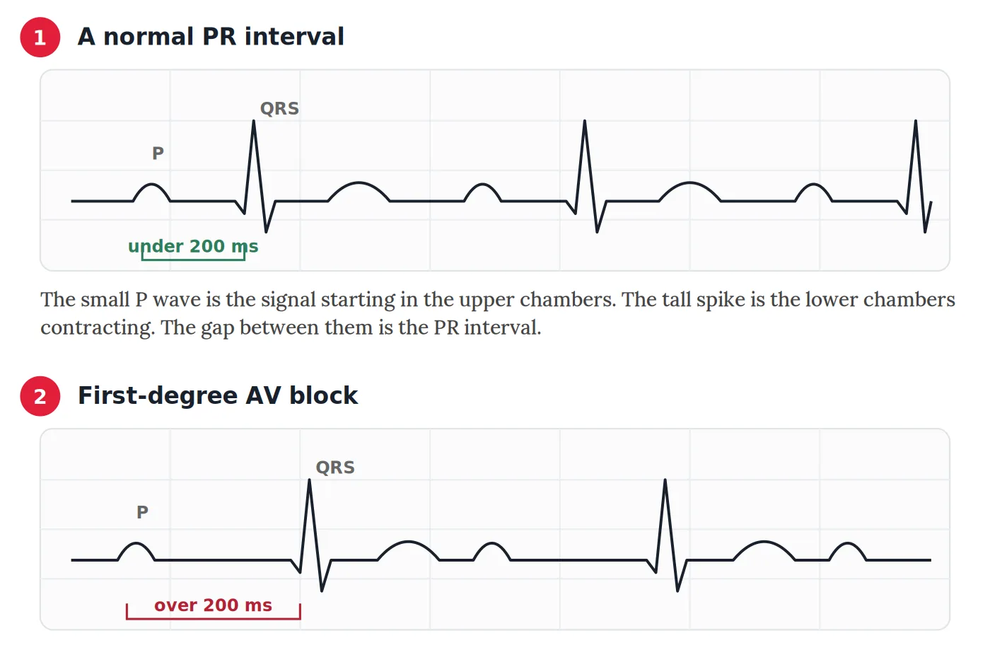
The crucial detail, and the one that separates this from the more significant conduction problems, is that every beat still gets through. In second-degree AV block some beats are dropped. In third-degree, or complete, heart block, the upper and lower chambers stop communicating altogether. Neither of those is what first-degree describes.
What Causes It
There are many reasons for a longer PR interval, and in a great many cases it does not indicate disease at all.
Normal variation. Some people simply have a conduction system that runs slightly slower than average, with no underlying problem and no consequence.
Being fit. This is one of the most common explanations and one of the most reassuring. Endurance athletes and very active people often have high vagal tone, meaning the nerve that slows the heart at rest is more dominant. The same physiology that gives an athlete a resting pulse in the 40s can lengthen the PR interval. It typically shortens again during exercise, which is one reason a stress test is sometimes used to sort this out.
Medications. Several common heart medicines slow conduction through the AV node deliberately, because that is part of how they work. Beta-blockers, certain calcium channel blockers, digoxin, and antiarrhythmic drugs can all lengthen the PR interval. When first-degree AV block appears in someone taking one of these, the medication is usually the explanation.
Ageing. The conduction system develops mild fibrous changes over the decades, much as other tissues do. A slightly longer PR interval at 75 is a fairly ordinary finding.
Underlying heart conditions. Less commonly, a prolonged PR interval forms part of a wider picture, particularly when the ECG shows other abnormalities alongside it. Ischaemic heart disease, inflammation of the heart muscle, and infiltrative conditions such as cardiac amyloidosis can all affect conduction. This is the minority, not the rule.
In an otherwise healthy person with no symptoms and an otherwise normal ECG, first-degree AV block is generally regarded as a benign finding.
When It Warrants a Closer Look
Most of the time, no further action follows. There are four situations where clinicians tend to look more carefully.
Symptoms. Dizziness, unusual tiredness, breathlessness on exertion, or fainting change the picture entirely. Those symptoms are not caused by first-degree AV block itself in most cases, but they raise the question of whether something more is going on alongside it.
A markedly long interval. Beyond about 300 milliseconds, the timing between the chambers can become inefficient enough to matter. The upper chambers may contract against closed valves, producing symptoms that resemble those of a poorly set pacemaker. This is uncommon.
Other findings on the same tracing. First-degree AV block appearing alongside a left bundle branch block or a right bundle branch block is a different proposition from first-degree AV block on its own, because it suggests the conduction system is affected at more than one level.
Family history. Inherited conduction disease is rare, but a family history of pacemakers at a young age, unexplained fainting, or sudden death in a relative is worth mentioning, because it changes how the finding is interpreted.
Does It Progress?
This is the question most people actually want answered, and the honest response is that it usually does not, but occasionally it can.
Large population studies have found that people with a prolonged PR interval have a somewhat higher long-term likelihood of developing atrial fibrillation, of needing a pacemaker, and of other conduction problems, compared with people whose PR interval is normal. The increase is measurable across a population but modest for any one person, and the majority never develop anything further.
That is the reason a repeat ECG is sometimes suggested at some point in the future rather than nothing at all. It is monitoring rather than treatment, and it reflects prudence rather than concern.
What Tests Might Follow
Where there is uncertainty, a small number of tests can clarify things.
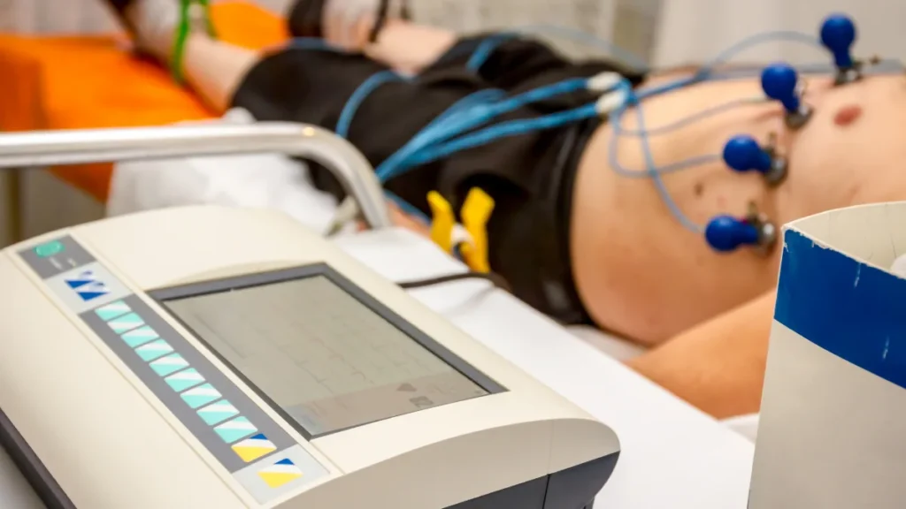
A repeat ECG shows whether the interval is stable or changing. A Holter monitor, worn for 24 hours or longer, catches intermittent conduction problems that a 10 second tracing in a clinic would miss. An echocardiogram looks at the structure and pumping function of the heart. And an exercise test can be informative, because a PR interval that shortens normally with exercise is reassuring, while one that lengthens is not.
Is Treatment Needed?
For the great majority of people, no treatment is required or recommended. First-degree AV block on its own, in someone without symptoms and with an otherwise normal heart, is generally left alone and observed.
Where a medication is responsible and the interval is long, the prescribing doctor may consider whether the dose still suits, weighed against whatever that medication was doing for the person in the first place. That is a judgement made case by case, not a rule.
A pacemaker is very rarely relevant here. It is reserved for the unusual combination of a markedly prolonged interval and genuine symptoms attributable to it, and it is not part of the usual course of this finding.
Conclusion
First-degree AV block is one of the most common findings on an ECG report and one of the most alarming-sounding. The name is the problem, not the condition. What it describes is a signal arriving slightly late, every time, in a heart that is otherwise doing exactly what it should.
If it turned up on a routine ECG and you feel well, the odds are strongly that it means very little. It is still worth asking your own doctor what it means in your particular case, especially if you take heart medications or the report mentioned anything else alongside it. Our heart glossary covers the other terms that tend to appear on the same page.
Related Reading
- The Electrocardiogram (ECG / EKG)
- What Does a Left Bundle Branch Block (LBBB) Mean on an ECG?
- Right Bundle Branch Block (RBBB): Why It Sounds Worse Than It Usually Is
- Ventricular Ectopic Beats: What They Are and What They Mean
- When a Slow Heart Rate Is Not Normal: Understanding Bradycardia
- The Holter Monitor: Catching Arrhythmias in Daily Life
- An Isolated Inferior Q Wave on Your ECG: Why It’s Usually Nothing to Worry About