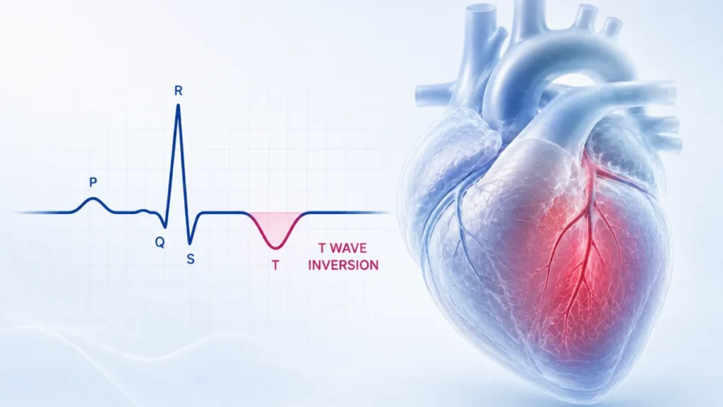
- An ECG records the heart from twelve different viewpoints. After each beat the heart resets, and that recovery shows on the tracing as a rounded wave called the T wave.
- In most viewpoints that wave points upwards. When it points downwards instead, the report describes it as T wave inversion.
- In several viewpoints a downward T wave is entirely normal and expected, so the phrase on its own says very little.
- What determines whether it matters is which viewpoints are involved, whether it is new compared with a previous tracing, and whether there are any symptoms.
- A previous ECG, if one exists, is often the single most useful piece of information, because a longstanding pattern and a new one carry very different weight.
Of all the phrases that turn up on an ECG report, T wave inversion is among the most common and the least self-explanatory. It sounds like something has been reversed, or has gone wrong in a way that ought to be obvious.
What it describes is narrower than that. A small wave on the tracing points downwards instead of upwards. In several of the heart’s twelve viewpoints that is completely normal. In others it can be important. The phrase alone does not tell you which situation you are in.
What the T Wave Is
Each heartbeat has two halves. The heart muscle activates and contracts, then it has to reset electrically before it can do it again.
On an ECG tracing, the tall spike is the contraction. The rounded wave that follows it is the reset. That second wave is the T wave.
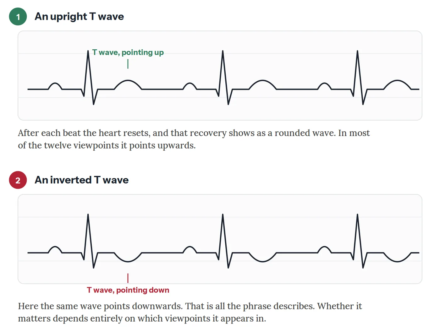
Because the twelve viewpoints look at the heart from different angles, the same electrical recovery appears differently in each. In most of them the T wave points upwards. In a few it normally points down, and always has.
Where a Downward T Wave Is Simply Normal
This is the part that resolves most people’s concern, and it is worth stating plainly.
One viewpoint, the one labelled aVR, has a downward T wave in essentially everybody. It looks at the heart from the opposite direction to the others, so an upward wave there would be the abnormal finding.
Two others, V1 and lead III, very commonly show a downward T wave in healthy people. So does lead V2 in some. An automated report will still flag these, because software counts rather than interprets.
There is also a pattern sometimes called the persistent juvenile pattern. Children normally have downward T waves across several of the chest viewpoints, and in most people these turn upward through adolescence. In some adults, more often women and more often people of African or Caribbean ancestry, the childhood pattern simply persists. It is a normal variant, not a disease.
In some viewpoints a downward T wave is the expected finding. The software flags it anyway, because it counts rather than interprets.
What Determines Whether It Matters
Three questions decide almost everything, and a cardiologist works through them in this order.
Usually needs nothing beyond an explanation
- The inversion sits only in aVR, V1, or lead III, where it is normal.
- An older tracing shows the same pattern, unchanged over years.
- There are no symptoms, and the rest of the ECG is normal.
- The person is young, fit, and the pattern fits a recognised normal variant.
Usually has a cause that is already known
- The inversion accompanies a bundle branch block, where changed conduction alters the recovery wave as a matter of course.
- It accompanies a thickened heart muscle, where the pattern is sometimes described as strain.
- Certain medicines, including digoxin, change the shape of the recovery wave predictably.
- Low potassium or magnesium can flatten or invert it, and correcting the level corrects the tracing.
The pattern, the symptoms, or the change over time raises concern
- The inversion is new compared with a previous tracing.
- It appears across several neighbouring viewpoints rather than one.
- It is deep and symmetrical rather than shallow, particularly across the front of the heart.
- There are symptoms, especially chest discomfort, breathlessness, or fainting.
The Pattern Cardiologists Look For First
There is one specific arrangement worth knowing about, because it is the reason this finding is never dismissed out of hand.
Deep, symmetrical T wave inversion across the viewpoints that look at the front of the heart, appearing in someone who has recently had chest pain that has since settled, can indicate a critical narrowing in the artery supplying that territory. The person often feels fine at the moment the tracing is taken, which is exactly what makes it dangerous.
It is uncommon. Most T wave inversion is nothing like this. But it is the reason a cardiologist will ask carefully about recent chest pain even when someone currently feels well, and why this particular pattern prompts prompt assessment of the coronary arteries rather than watchful waiting.
Other Causes Worth Knowing
Beyond the coronary arteries, several conditions change the recovery wave.
A previous heart attack leaves permanent changes in the viewpoints that looked at the damaged area, often alongside Q waves.
Thickened heart muscle, whether from long-standing high blood pressure or from hypertrophic cardiomyopathy, alters recovery across several viewpoints. In the inherited condition the changes can be striking, and they are one of the findings that prompts further imaging in a young person.
Inflammation of the heart muscle or its surrounding sac produces widespread changes rather than ones confined to a single artery’s territory.
A clot in the lungs can invert the T waves across the right-sided viewpoints, usually alongside breathlessness and a fast heart rate.
Conditions affecting the brain, including bleeding around it, can produce dramatic deep T wave inversion. These are sometimes called cerebral T waves, and the heart itself is normal.
Athletic training changes the ECG in ways that overlap with disease, and distinguishing the two is a recognised specialty in its own right. Ancestry matters here: T wave inversion across the front chest viewpoints is a well-described normal finding in athletes of African or Caribbean ancestry, and considerably more concerning in others.
What Usually Happens Next
The single most useful step is finding an old ECG. A pattern that has been present for a decade means something entirely different from one that appeared last month, and comparison answers the question faster than any new test.
If there is no previous tracing, or the pattern is new, the usual next steps depend on the picture. A troponin blood test checks whether heart muscle has been damaged. An echocardiogram looks at the muscle thickness and how well each area is moving, which can show a problem in the same territory the ECG pointed at.
Where the coronary arteries are the concern, a stress test or a CT coronary angiogram follows. Where the finding is unexplained in a young person, cardiac MRI is often used, because it shows the muscle in more detail than ultrasound can.
And in a great many cases none of this is needed, because the inversion sits in a viewpoint where it belongs and the conversation ends there.
Conclusion
T wave inversion describes the direction of a small wave on a tracing. That is genuinely all the phrase means, and in several viewpoints the downward direction is the normal one.
What turns it into something worth investigating is context: which viewpoints, whether it is new, and whether anything else is going on. Those are questions a person cannot answer from the report alone, and they are the right things to ask.
If you have been given this finding, two pieces of information will shape the conversation more than anything else. Whether an older ECG exists for comparison, and whether you have had any symptoms, particularly chest discomfort, in the weeks beforehand. Both are worth mentioning without being asked.
Related Reading
- The Electrocardiogram (ECG / EKG)
- An Isolated Q Wave on Your ECG: Why It’s Usually Nothing to Worry About
- What Does a Left Bundle Branch Block (LBBB) Mean on an ECG?
- Right Bundle Branch Block (RBBB): Why It Sounds Worse Than It Usually Is
- First-Degree AV Block on Your ECG: What Does It Really Mean?
- Hypertrophic Cardiomyopathy: What HCM Actually Means
- The Echocardiogram: What It Shows and Why It Matters