The Pulse Newsletter Expert heart health, in plain English. Fortnightly, from our clinicians. Subscribe →

Cardiac Rehabilitation: The Heart Treatment Most People Miss

cardiac rehabilittion
Key Points

  • Cardiac rehabilitation is a structured program of supervised exercise, education, and emotional support offered after a heart attack, a stent procedure, bypass surgery, and several other heart conditions.
  • A Cochrane review of 85 trials involving more than 23,000 people found that exercise-based rehabilitation lowered the chance of another heart attack and of being admitted to hospital in the following year.
  • Supervised exercise after a heart event is very safe. One large study recorded about one serious cardiac event for every 50,000 hours of exercise training.
  • Only around 4 in 10 eligible people take part, and women and older people are the least likely to be referred or to attend.
  • Trials found similar benefits whether rehabilitation took place at a centre or at home, which opens the door for people who live far away, work, or care for others.

Leaving hospital after a heart attack or heart surgery can feel like being handed back a body that no longer feels familiar. Walking to the letterbox raises questions. So does climbing stairs, going back to work, or simply sleeping through the night without worry.

Cardiac rehabilitation exists to answer those questions. It is one of the best-studied treatments in cardiology, yet it is also one of the most underused. Many people who would benefit never start, and many who start do not finish.

What Is Cardiac Rehabilitation?

Cardiac rehabilitation, often shortened to cardiac rehab, is a coordinated program that helps people recover after a heart event and lowers the chance of another one. It combines supervised exercise with education about the heart, medicines, and lifestyle, along with support for the emotional side of recovery.

It is not a single class or a leaflet. It is usually run by a team that can include nurses, exercise physiologists, physiotherapists, dietitians, psychologists, and doctors. The exercise is tailored to each person’s fitness and condition, then increased gradually as confidence and strength return.

Who It Is For

Rehabilitation is most often offered to people who have had a heart attack, a stent inserted during a coronary angiogram, or bypass surgery. People living with stable angina are also commonly referred.

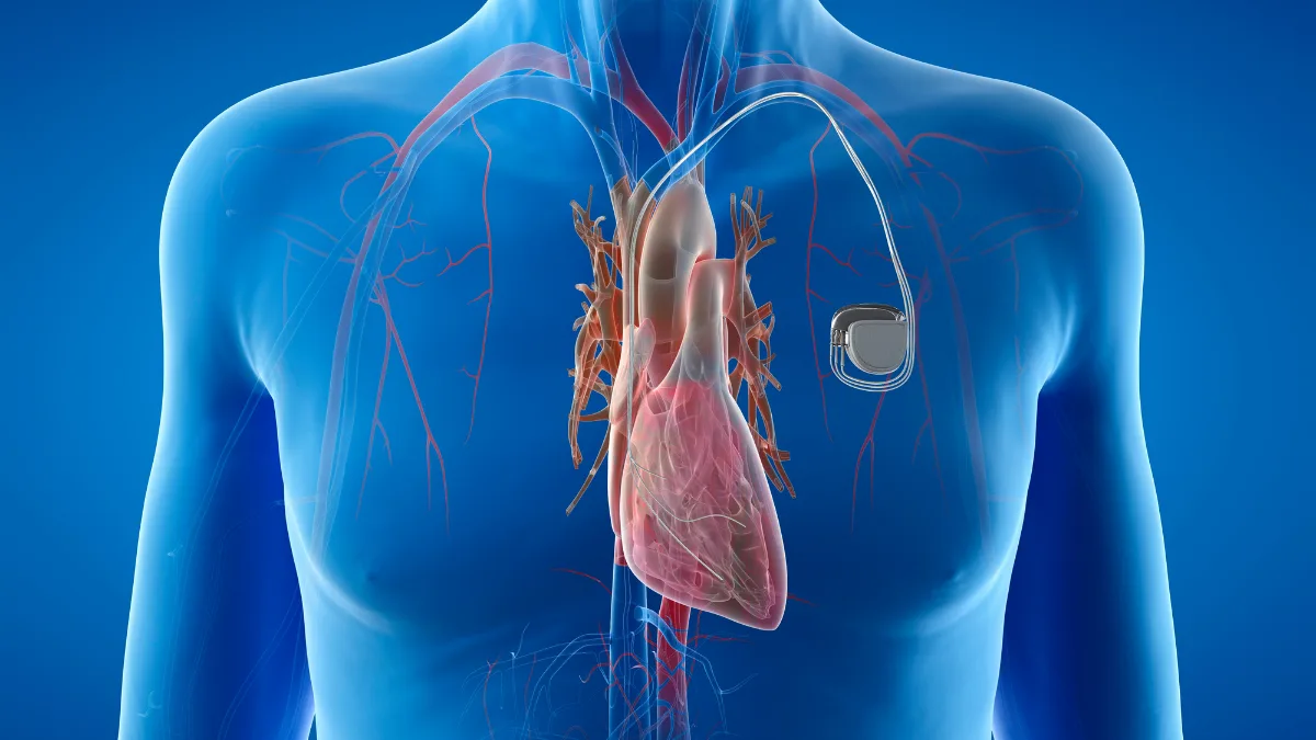
Many programs also accept people with heart failure, people recovering from heart valve surgery, and people who have had a pacemaker or defibrillator fitted. Which conditions qualify varies between countries and services. Whether a program suits a particular person is worth discussing with their cardiologist or treating team.

The Three Phases of Rehabilitation

Rehabilitation is often described in phases. The names and numbering differ between countries, but the overall journey is broadly similar everywhere.

1
In hospital
Recovery begins on the ward. Staff help people get up and walk safely, explain what has happened to the heart, and arrange referral to an outpatient program before discharge.

2
Structured program
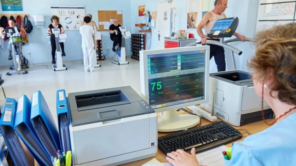
The core of rehabilitation. Supervised exercise sessions, usually one to three times a week over several weeks, run alongside education and support. This can take place at a hospital, a community centre, or at home.

3
Lifelong maintenance
The habits built during the program carry on independently. Some people join community exercise groups, while others continue on their own with occasional check-ins.

What Happens in a Program

Programs usually begin with an assessment. This usually covers symptoms, medicines, fitness, risk factors such as blood pressure and cholesterol, and how the person is coping emotionally. The results shape a plan that fits the individual.

What a program usually includes
Aerobic exercise
Walking, cycling, or circuit work, monitored by staff and built up gradually.

Strength work
Light weights or resistance bands to rebuild muscle and everyday strength.

Heart education
How the heart works, what happened, and which symptoms need attention.

Medicines
What each medicine does and why it has been prescribed.

Emotional support
Help with anxiety, low mood, and the loss of confidence common after a heart event.

Lifestyle
Support with eating well, stopping smoking, sleep, and returning to work and driving.

The exercise sessions usually centre on activities such as walking and stationary cycling. Many programs also include resistance training, which helps with everyday tasks such as carrying shopping or getting up from a chair.

The education sessions often cover eating patterns such as the Mediterranean diet, and practical support for stopping smoking. Family members and carers are often welcome to attend.

What the Evidence Shows

The most thorough look at the evidence is a Cochrane review that pooled 85 randomised trials involving 23,430 people with coronary heart disease. Most had had a heart attack, a stent, or bypass surgery. The review compared people who took part in exercise-based rehabilitation with people who received usual care without structured exercise.

12 in 100
people in rehabilitation were admitted to hospital within a year, compared with 21 in 100 who did not take part.
Cochrane Review, 85 trials
1 in 75
For every 75 people who took part, one heart attack was prevented within a year.
Cochrane Review, 85 trials

In plain numbers, about 5 in 100 people who did not take part had a heart attack within a year, compared with between 3 and 4 in 100 of those who did. The reviewers rated this finding as high certainty. The fall in hospital admissions was rated moderate certainty.

The effect on survival within the first year was small and less certain, at about 5 in 100 deaths with rehabilitation compared with about 6 in 100 without. Trials that followed people for longer found fewer deaths from heart causes among those who took part, although fewer trials contributed to that result.

Quality of life scores also improved slightly, although the differences may be too small for many people to notice. The benefits were broadly similar regardless of the type of heart condition, whether the program was exercise alone or combined with education, and where it took place.

The evidence has limits. Most people in the trials were middle-aged men, and women made up fewer than 15% of participants. Findings in women and in older people with several health conditions rest on thinner data.

Rehabilitation is not a reward for recovering well. It is part of how recovery happens.

Is Exercise Safe After a Heart Event?

Fear of bringing on another heart attack is one of the most common reasons people hold back. The data are reassuring. One large study of more than 25,000 people in rehabilitation programs recorded about one serious cardiac event for every 50,000 hours of exercise training.

Part of that safety comes from the assessment at the start, which sets exercise at a level suited to the individual. Staff also watch for chest pain, unusual breathlessness, dizziness, or palpitations during sessions. These symptoms usually prompt a pause in exercise and a medical review.

Why So Many People Miss Out

Despite strong guideline support from the European Society of Cardiology and the American Heart Association, only around 4 in 10 eligible people take part. Some are never referred. Others are referred but do not start, or start but do not finish.

The reasons are often practical. Travel distance, work commitments, caring responsibilities, and cost all play a part, and access and funding vary widely between countries. Some people feel well after a stent and see no need for it. Others feel too anxious or unwell to begin.

Women and older people are the least likely to be referred and to attend. This matters because both groups face a real risk of further heart problems, and both stand to gain from the support rehabilitation provides, as outlined in recovering after a heart attack.

Home-Based and Virtual Programs

Rehabilitation does not have to take place in a hospital gym. In the Cochrane review, about a quarter of the trials delivered rehabilitation entirely at home, some using mobile phones or the internet. The benefits were similar regardless of setting.

Home and virtual options can suit people in rural areas, people who work, and people who are uncomfortable in group settings. Centre-based programs offer direct supervision and the encouragement of others on the same path. The format that works best is the one a person can keep attending, and the choice is typically made together with the rehabilitation team.

Conclusion

Cardiac rehabilitation lowers the chance of another heart attack and of returning to hospital, and it helps people regain strength and confidence after a frightening event. It is safe, well supported by evidence, and offered in several formats, yet most eligible people never take part.

For anyone recovering from a heart attack, a stent, or heart surgery, rehabilitation is worth discussing with the treating cardiologist or hospital team. It is one of the most useful conversations in the whole recovery journey.

Related Reading

What a Pacemaker Does, and What Life With One Is Like

heartmatters.com 90

Being told you need a pacemaker can feel like a significant moment. In practice it is one of the most established procedures in cardiology, and for a heart beating too slowly the difference it makes is usually large and quick.

Five Ways to Get More Out of Your Air Fryer

air fryer

Key Points

  • An air fryer is a small, very fast convection oven, and it earns its place in a heart-healthy kitchen mainly through what it makes easy rather than what it removes.
  • Vegetables are what it does best. Roasting them takes around 12 minutes and needs barely any oil, which turns a side dish into something people actually look forward to.
  • A teaspoon of olive oil brushed on goes further than a long spray, and extra virgin olive oil handles the temperatures most air fryers run at.
  • Oily fish cooks in under 10 minutes with no pan and no added fat, which makes the twice-weekly fish habit considerably less effortful.
  • Herbs, spices, citrus, and vinegar carry most of the flavour that salt usually carries, and the seasoning step is where a meal’s sodium content is decided.

The air fryer arrived in most kitchens as a chip machine. That is a shame, because chips are close to the least interesting thing it does.

What it is really good at is dry, fast, high heat in a small space, which happens to be exactly what vegetables, legumes, and fish respond well to. Used that way, it quietly removes several of the reasons people give for not cooking the food they already know they should be eating. Here are five things worth knowing.

One: Vegetables Are What It Does Best

Roasted vegetables are one of the great sells of Mediterranean-style eating, and the usual barrier is the oven. Preheating a large oven for 40 minutes to roast a tray of broccoli is a poor trade, and most people steam them instead and find the result forgettable.

An air fryer needs no meaningful preheat and works in about 12 minutes. Broccoli, cauliflower, carrots, capsicum, Brussels sprouts, and pumpkin all crisp at the edges and sweeten in the middle, which is a different food from the boiled version. Chickpeas from a tin, drained and dried, come out crunchy in around 15 minutes and make a snack with real fibre in it, which is one of the more underrated dietary influences on cholesterol.

This matters more than it sounds. The gap between knowing vegetables are good and eating them is usually about effort, not knowledge.

Two: A Refillable Mister Is the Best Thing in the Drawer

Most air fryer recipes call for a light spray, and most people reach for an aerosol can to provide it. A refillable pump mister filled with olive oil does the same job better, and it is probably the single most useful item to sit next to an air fryer.

Aerosol cooking sprays contain propellants, anti-foaming agents, and emulsifiers alongside the oil, and over time they tend to leave a gummy residue that dulls the non-stick coating on the basket. A pump mister holds nothing but oil. It delivers a genuinely fine mist, so a vegetable tray is coated with a fraction of a teaspoon rather than the tablespoon a bottle would pour, and the bottle is refilled from whatever oil is already in the kitchen.

Extra virgin olive oil is the sound choice to fill it with. Most air fryers run between 160 and 200°C, which sits within the range extra virgin olive oil tolerates comfortably, and the flavour it carries means less seasoning is needed later. Misting the food rather than the basket is the detail that matters, since oil sprayed onto bare metal burns onto the surface rather than doing anything useful.

The most useful thing in my kitchen for the air fryer is a refill spray bottle of olive oil. It costs almost nothing and it changes how much oil you actually use.

Prof. Peter Barlis, Interventional Cardiologist

Three: Oily Fish, Under 10 Minutes, No Pan

The advice to eat fish twice a week is among the most widely agreed-upon pieces of dietary guidance in cardiology, and among the least followed. The reasons people give are consistent: the smell, the pan, the washing up, and the fear of overcooking it.

An air fryer removes three of those four. A salmon fillet takes roughly eight to nine minutes at 190°C, needs no added fat because the fish provides its own, and the enclosed basket contains most of the aroma. Our air fryer salmon with pumpkin chips and broccoli cooks the whole plate in one basket, and the reasons salmon earns its reputation have nothing to do with the appliance and everything to do with what is in the fish.

Sardines, mackerel, and trout behave similarly, and are generally cheaper.

Four: Seasoning Is Where the Sodium Is Decided

The cooking method has no effect at all on how much salt ends up in a meal. That is decided in the 10 seconds before the basket closes, and it is the single most useful habit to think about in an air fryer kitchen.

Dry heat concentrates flavour, which means the food needs less help than it would if boiled. Smoked paprika, cumin, garlic powder, dried oregano, chilli flakes, and black pepper all bloom in the hot air. A squeeze of lemon or a splash of red wine vinegar after cooking does much of what salt does to brighten a dish, without the sodium. Since dietary sodium is one of the more consistent influences on blood pressure, this is worth more attention than most air fryer content gives it, and it sits alongside the other practical influences on blood pressure.

The other half of the sodium question is what goes in the basket in the first place. Frozen crumbed products carry their salt with them regardless of how they are cooked.

Five: Gold, Not Brown

Acrylamide is a compound that forms when starchy foods such as potato and bread brown at high temperature. It comes from heat rather than oil, so an air fryer has no automatic advantage over any other high-heat method, and the published comparisons between cooking methods do not agree with each other.

What does show up consistently is that darker browning means more acrylamide. Cooking potato to a light gold rather than a deep brown is the practical version of that finding. Soaking cut potato in cold water for half an hour before cooking, then drying it, also lowers acrylamide levels, and has the useful side effect of producing a crisper result.

None of this applies to vegetables, fish, or meat, which do not form acrylamide in any meaningful quantity. It is a starch and high heat phenomenon.

Vegetables and legumes

Broccoli, cauliflower, carrots, capsicum, pumpkin, and tinned chickpeas. Roughly 12 to 15 minutes at 190°C, a teaspoon of olive oil, single layer in the basket.

Oily fish

Salmon, trout, mackerel, and sardines. Eight to nine minutes at 190°C, no added oil needed, skin side down. The enclosed basket keeps most of the aroma in.

Flavour without salt

Smoked paprika, cumin, oregano, garlic, chilli, and pepper before cooking. Lemon juice or vinegar after. Dry heat concentrates flavour, so less seasoning is needed overall.

The One Thing It Does Not Do

An air fryer changes how food is cooked. It has no effect on what goes into it. Frozen crumbed chicken pieces come out of the basket with the same salt, refined starch, and additives they had in the packet, and a lower-fat cooking method is not a reason for a larger serving.

The appliance is a good servant of a Mediterranean pattern of eating and a poor substitute for one. Fill the basket with vegetables, fish, and legumes and it is genuinely useful. Fill it with the frozen aisle and it is a faster version of the same meal.

Conclusion

The reason an air fryer is worth having is not that it removes fat, though it does that when it replaces deep frying. It is that it shortens the distance between a raw vegetable and a dinner someone wants to eat, and most of the difficulty in eating well sits in that distance.

A basket of roasted vegetables, a fillet of oily fish, a teaspoon of olive oil, and a decent spice rack will cover most weeknights. The machine is incidental. What goes in it is not.

Related Reading

Cholesterol After Menopause: What Changes and When

menopause cholesterol
Key Points

  • The cholesterol rise around menopause is driven by the ovaries winding down, not by getting a year older. It appears over a short window rather than drifting slowly across midlife.
  • Studies that followed the same women year after year found LDL cholesterol climbed sharply within about a year either side of the final period, then levelled off at the higher level.
  • HDL cholesterol is the misleading part. The number often holds steady or rises during this time, while the particles themselves change in ways that make them work less well.
  • Women whose LDL rose fastest across this window were more likely to have plaque in their neck arteries years later than women whose LDL rose more slowly.
  • An earlier menopause means the altered cholesterol pattern starts sooner and lasts longer.

Cholesterol is usually described as something that creeps upward with age, the same way for everyone. For women going through menopause, that description misses something important. The change is not a slow creep. It is not about getting older.

It is a distinct shift, and it clusters around one point in time. It is also easy to miss, because the years in which it happens are crowded with symptoms that feel far more pressing.

What Actually Changes

Oestrogen affects how the liver handles cholesterol. It helps keep active the docking points on liver cells that pull LDL particles out of the bloodstream. As oestrogen falls, that removal slows down, and LDL levels rise.

Total cholesterol rises alongside it. So does a measure called apolipoprotein B, usually shortened to apoB. Rather than measuring how much cholesterol is being carried, apoB counts how many of the particles that lodge in artery walls are in the blood. That count often tells more than an LDL figure on its own.

Body shape tends to change over the same years, with more fat settling around the middle. That affects how the body handles both cholesterol and blood sugar, and it adds to the picture.

The Window Around the Final Period

The most useful research followed the same women repeatedly across the transition. That design answers a question earlier studies could not: whether the cholesterol changes belong to the ovaries winding down, or to the calendar.

The answer is reasonably clear. LDL cholesterol and apoB rise sharply within about a year either side of the final period. They then settle at the higher level rather than continuing to climb. That is not the pattern ageing produces on its own.

Not everything on a blood test behaves this way. Triglycerides, blood pressure, and blood sugar also tend to drift upward through midlife. They follow the slower line of age rather than clustering around the final period. The same menopause pattern appeared in every ethnic group studied.

How big is the change?

Small, in any single year. That is much of the reason it goes unnoticed.

One study looked at how fast LDL climbed across this window and then scanned the same women’s neck arteries more than a decade later. Women in the fastest-rising third, gaining roughly 0.19 mmol/L (about 7 mg/dL) a year or more, were about half as likely again to have plaque in those arteries as women in the slowest-rising third, who gained under about 0.14 mmol/L (5 mg/dL) a year.

The gap between those two groups is not dramatic on a single blood test. It is the kind of difference that would be easy to dismiss as noise. What made it visible was measuring the same women repeatedly, and this is an observed link rather than proof that the cholesterol change caused the plaque.

Rises sharply

LDL cholesterol, total cholesterol, and apoB. The climb sits in the year before and after the final period, then levels off.

Looks unchanged, but is not

HDL cholesterol often holds steady or rises. Underneath, larger particles decline, smaller ones increase, and each one clears less cholesterol.

Carries forward

Cholesterol changes measured across this window have been linked to thickening and plaque in the neck arteries years afterwards.

The HDL Number That Misleads

This part deserves more attention than it gets, because an ordinary blood test can offer false reassurance.

HDL cholesterol has long been treated as the good number. Across the menopause transition it often stays level or even rises. Taken at face value, that reads as encouraging news.

Looking at the particles themselves tells a different story. In the couple of years around the final period, the larger HDL particles decline and the average particle gets smaller. The number of small particles rises. Each particle also becomes less effective at collecting cholesterol out of tissue and carrying it away. More particles, each doing less.

A steady HDL number through menopause is not proof that nothing has changed. It is one of the few figures on the panel that can move in the wrong direction while appearing not to move at all.

The practical consequence is that a reassuring HDL result does not cancel out a rising LDL or apoB when a test from these years is being interpreted.

Why This Gets Missed

Several things converge. The transition brings symptoms that are immediate and disruptive. A cholesterol test competes poorly for attention against broken sleep, hot flushes, night sweats, and low mood.

Heart risk assessment in women has also historically happened less often and later than in men. That pattern is examined further in the article on why women’s heart health is different.

Risk calculators add their own problem. Most lean heavily on age. A woman in her early 50s can return a low 10-year risk score at exactly the moment her cholesterol is changing fastest. The score is not wrong on its own terms. It is answering a question about the next decade, not about the direction of travel.

Timing matters too. A woman whose last period comes in her mid-40s spends more years with the altered pattern than one who reaches it at 55. A calculator reading age alone does not capture that.

What Can Be Measured

A cholesterol test taken during the transition says more when there is an earlier one to compare it against. The direction of change carries information a single figure does not. This is why the researchers behind the artery findings suggested that checking cholesterol more often through perimenopause may be worth considering. Where apoB is available, it adds the particle count to the picture.

Lipoprotein(a) is largely inherited and is usually measured once in a lifetime rather than tracked. It does not appear to change with menopause. It is worth knowing about all the same, because it is a test many people never have at all, and the transition is one of the points at which it tends to come up.

Sometimes a risk score still leaves real uncertainty about what the arteries are doing. Several further tests exist. Whether any of them is worth considering, and which one, depends entirely on the individual picture, and that is a decision made with a doctor who knows the full history.

More testing is not automatically better. Each test has limitations as well as uses, none is used routinely, and guidance on who they suit differs from one country to the next. Heart Matters covers the coronary calcium score separately for readers who want to understand what one of these tests involves.

Where Hormone Therapy Fits

This is the question that comes up most often, and the answer has two parts that are easy to run together.

Hormone therapy does change cholesterol. It lowers LDL. Tablets also raise triglycerides in a way that patches and gels largely do not, which is one reason the two forms are not interchangeable. Tablets pass through the liver before reaching the bloodstream, and the liver is where most cholesterol handling happens.

The second part is the one that gets lost. Hormone therapy is not prescribed to prevent heart disease. Current guidance does not support using it for that purpose, and a better cholesterol reading on its own is not treated as a reason to start it.

That is not the same as saying it is off the table. Where hormone therapy is being considered for menopausal symptoms, its effect on cholesterol is one factor weighed within that decision, alongside age, time since the final period, and personal and family history. The wider discussion of timing and heart risk sits in the women’s heart health article.

What Else Helps

Nothing here is specific to menopause, but it all carries more weight when LDL is rising. Beta-glucan, the soluble fibre in oats, has consistent evidence for lowering LDL at amounts achievable in an ordinary breakfast. Phytosterols work a different way, by reducing how much cholesterol the gut absorbs.

Resistance training deserves a specific mention through these years. The loss of muscle that comes with the transition affects how the body handles cholesterol and blood sugar as well as bone strength, and aerobic exercise on its own addresses that less well.

Where cholesterol levels warrant medication, there is a historical point worth knowing. Statins have been prescribed to women less often, and at lower doses, than to men with comparable risk, despite the trial evidence supporting their use. The questions women raise most often are covered in the article on what patients ask most about statins.

What These Conversations Usually Cover

Cholesterol during menopause is not a single number to be judged pass or fail. These are the three things that tend to shape the discussion.

The direction, not just the number

Whether an earlier result exists to compare against. A figure inside the normal range can still be moving quickly.

When menopause happened

The age of the final period sets how many years the altered pattern has been running. Most risk scores do not ask.

What the score is measuring

A 10-year risk score answers a question about the next decade. It is not designed to flag a change happening now.

Heart Matters Resource

When in Doubt, Get Checked Out

Cholesterol changes quietly and causes no symptoms. Chest discomfort, unusual breathlessness, and unexplained fatigue are a different matter entirely, and in women they are more often put down to something else first.

Read: When in Doubt, Get Checked Out →

Conclusion

The cholesterol changes of menopause are not dramatic in any single reading. That is much of the reason they pass unremarked. They amount to a modest rise in LDL, a modest rise in apoB, and an HDL figure offering reassurance it has not entirely earned.

What gives them weight is the link between changes measured across this window and the state of the arteries years afterwards. Menopause is not only a set of symptoms to be managed. It is also a point at which the long-term picture shifts, and one of the few moments when a blood test taken at the right time says more than the same test taken at any other.

Related Reading

Left Ventricular Hypertrophy (LVH) on Your ECG

LVH on Your ECG: What Left Ventricular Hypertrophy Means
Key Points

  • “Left ventricular hypertrophy” on an ECG report means the tracing showed larger than usual electrical voltages, which can happen when the heart’s main pumping chamber has thickened. It is a suggestion, not a diagnosis.
  • The ECG measures electrical voltage rather than muscle thickness. It picks up a great deal well, but wall thickness is inferred rather than measured, so a normal ECG does not rule thickening out.
  • Body build affects the reading. Where there is less tissue between the heart and the electrodes, the voltages run higher with no thickening at all.
  • An echocardiogram settles the question, because it measures the muscle directly rather than inferring it from voltage.
  • Where genuine thickening is confirmed, the cause matters more than the finding. High blood pressure is by far the most common, and the thickening can partly reverse when the pressure is treated.

Among the phrases that automated ECG software generates, “left ventricular hypertrophy” is one of the more alarming to read. It sounds like a diagnosis of something structural and permanent. Hypertrophy means enlargement. Left ventricular means the main pumping chamber. Put together, it reads as though a machine has just told you your heart is abnormally big.

What the machine has actually done is add up some voltages and compare the total against a threshold. Whether that reflects anything real about your heart is a separate question, and one the ECG is not well equipped to answer.

What the Left Ventricle Does and Why It Thickens

The left ventricle is the chamber that pushes blood out to the entire body. It is the thickest-walled part of the heart because it does the most work.

Like any muscle, it responds to sustained extra load by getting thicker. If it has to push against higher pressure with every single beat, year after year, the wall builds up in response. That is left ventricular hypertrophy.

It is not a disease in itself. It is an adaptation, and the important question is always what it is adapting to.

What the ECG Can and Cannot Tell You

The ECG is a genuinely valuable test, and it is ordered as often as it is for good reason. It detects rhythm disturbances, conduction problems, and the changes of a heart attack quickly, cheaply and reliably. For those questions it is hard to better.

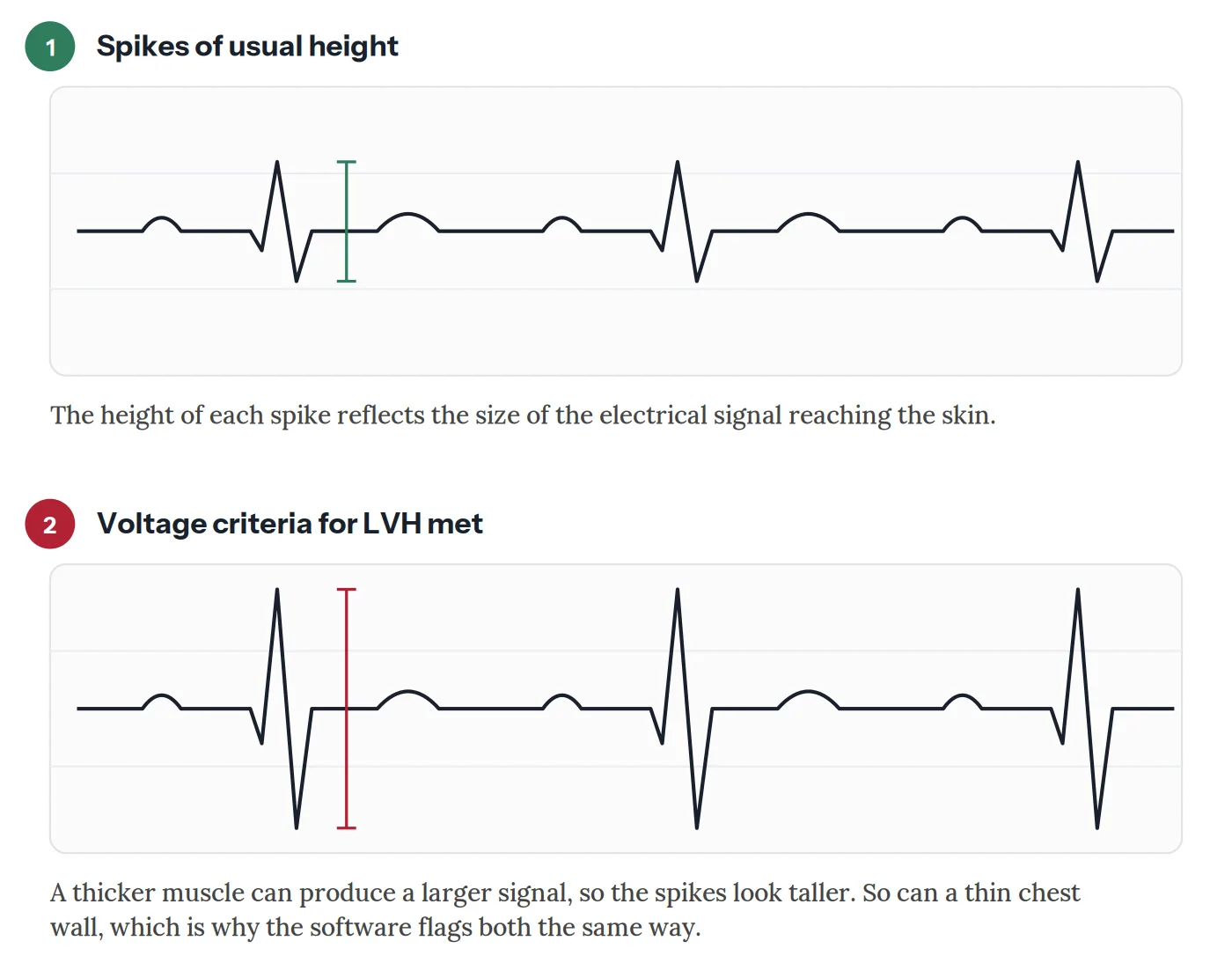
Wall thickness is simply not one of the things it measures directly. It measures electrical voltage at the skin. The reasoning behind the criteria is that a thicker muscle generates a larger electrical signal, so an unusually large signal might indicate a thicker wall.

That reasoning is sound, but voltage is an indirect measure. Depending on which criteria are used and which population is studied, the ECG identifies somewhere between roughly a fifth and a half of people who genuinely have a thickened left ventricle on imaging. It is a screening signal rather than a measurement.

The reason is that voltage at the skin depends on a great deal besides the heart. Chest wall thickness, body size, lung volume, the position of the heart in the chest, and even the exact placement of the electrodes all influence the reading.

A thicker muscle and a thinner chest wall both raise the voltage, which is why the ECG raises the question rather than answering it.

This is why the finding turns up so often in people whose hearts are entirely normal. Where there is less tissue between the heart and the electrodes, the signal arrives stronger and the voltages read higher. Regular endurance training adds to this, partly for the same reason and partly because it produces some genuine and entirely healthy adaptation of the muscle.

The reverse happens too. Someone carrying extra weight, or with lung disease, may have a genuinely thickened ventricle and a completely unremarkable ECG, because the signal is dampened before it reaches the skin. This is why a normal tracing does not settle the question either.

Two ECG tracings compared. In the first the spikes are of usual height. In the second they are markedly taller, which is what an automated report means by voltage criteria for left ventricular hypertrophy
A thicker muscle can produce a larger signal, so the spikes look taller. A thin chest wall can do the same, which is why the finding prompts a closer look rather than settling the question, Heart Matters

What the Report Might Say

The wording varies between machines, and the differences carry meaning.

“Voltage criteria for LVH” is the mildest version. It means only that the numbers exceeded a threshold. In someone with no symptoms and nothing else on the tracing, this on its own very often means nothing.

“LVH with repolarisation abnormality”, sometimes written as a strain pattern, is a more meaningful finding. Here the tracing shows not just large voltages but changes in how the heart muscle recovers between beats. That combination is more likely to reflect genuine thickening and is more strongly linked to cardiovascular risk over time.

“Left atrial enlargement” appearing alongside it adds weight too, because the upper chamber often enlarges in response to a stiff, thickened ventricle below it.

What Causes Genuine Thickening

High blood pressure is by far the most common cause and the reason the finding matters. Every beat against elevated pressure asks more of the muscle, and the wall thickens accordingly. This is often the first hint that blood pressure has been higher than realised for longer than realised.

Aortic stenosis, where the valve at the exit of the left ventricle narrows, creates the same problem more acutely. The chamber has to force blood through a restricted opening, and thickens in response. Aortic stenosis is usually audible as a murmur, so it rarely comes as a surprise on an ECG alone.

Athletic training produces genuine adaptation in endurance and strength athletes, and it is generally benign and reversible with detraining. Distinguishing it from the conditions below is one of the more nuanced judgements in cardiology.

Hypertrophic cardiomyopathy is an inherited condition in which the muscle thickens without any load driving it. It is much less common than the causes above, but it matters because it can affect young people and runs in families. HCM is one reason an unexplained finding in a young person is worth following up rather than dismissing.

Infiltrative conditions such as cardiac amyloidosis can thicken the wall with deposited protein rather than muscle. A distinctive combination sometimes appears here: a wall that looks thick on imaging alongside unexpectedly small voltages on the ECG.

What Happens Next

The finding is usually followed by an echocardiogram, an ultrasound that measures the wall thickness directly and calculates the muscle mass. It answers in twenty minutes what the ECG could only infer, and it also shows whether the chamber is pumping and filling normally.

Watch: what an echocardiogram involves, what it measures, and why it answers questions the ECG can only raise, Heart Matters Educational Series

Blood pressure assessment usually follows too, and often means readings taken at home or over 24 hours rather than a single clinic measurement, since a one-off reading is a poor guide to the pressure the heart has actually been working against.

Where thickening is confirmed and the cause is not obvious, further tests may follow depending on the pattern seen, which might include cardiac MRI, blood tests, or a family history and genetic assessment if an inherited condition is suspected.

Why It Is Worth Knowing About

Confirmed left ventricular hypertrophy is not something to panic about, but it is not nothing either. It is associated over the long term with a higher likelihood of atrial fibrillation, of heart failure, and of stroke, which is why cardiologists take it seriously as a marker even when the person feels entirely well.

The encouraging part is that it is not necessarily permanent. Where high blood pressure is the cause, sustained treatment can produce measurable regression of the thickening over months to years. The muscle that built up in response to pressure can partly unbuild when the pressure comes down. Studies have found that people whose ECG signs of hypertrophy regress with treatment go on to have fewer cardiovascular events than those whose do not.

That makes this one of the more actionable findings on an ECG report. It points at something that can be changed.

Conclusion

“Left ventricular hypertrophy” on an automated ECG report is a prompt to look further, not a verdict. In someone with no symptoms and an otherwise normal tracing it very often reflects nothing more than how the signal reaches the skin. In someone with high blood pressure it may be the first visible sign of what that pressure has been doing quietly for years.

Either way, the next step is the test that measures the muscle directly. An echocardiogram does that, and it is straightforward. What matters most is the conversation about what might be driving it, because for the most common cause, treatment does not just stop the thickening progressing. It can reverse some of it.

Related Reading