The Pulse Newsletter Expert heart health, in plain English. Fortnightly, from our clinicians. Subscribe →

Possible Old Heart Attack on ECG? What Q Waves Mean

heartmatters.com 2026 03 31T225746.389
Key Points

  • Receiving an ECG report that mentions “possible old heart attack” or “cannot exclude prior infarction” is one of the most anxiety-provoking findings in cardiology, and in an otherwise fit, healthy person with no symptoms, it is almost always a normal variant that requires no treatment.
  • An ECG records the heart’s electrical activity from 12 different viewpoints around the chest and limbs. A Q wave is simply a small downward dip seen in some of these viewpoints. In certain positions, particularly one called lead III, a Q wave is extremely common in completely normal, healthy hearts.
  • Automated ECG software flags Q waves as “possible old heart attack” because it cannot apply clinical judgement. A cardiologist reviewing the same ECG in the context of a fit, symptom-free person will almost always reach a very different and reassuring conclusion.
  • A Q wave that genuinely indicates a previous heart attack must appear in multiple neighbouring viewpoints simultaneously and be accompanied by other supporting changes on the ECG. An isolated Q wave in a single viewpoint, with everything else completely normal, does not meet this standard.
  • If you have received a report like this and are anxious, that anxiety is entirely understandable. A clinical review with a cardiologist resolves this question quickly and in most cases definitively.

One of the most common referrals I receive, easily once or twice a week, is a fit, healthy, often younger person who has had an ECG for a routine reason and whose report has come back with a phrase like “possible old heart attack,” “Q waves, cannot exclude prior infarction,” or “inferior changes, clinical review advised.”

They arrive in my clinic worried. Some have been unable to sleep. Some have told their families. Some have already started researching heart attack symptoms they have never had. The phrase “old heart attack” in a document about their heart has landed with enormous weight.

In the overwhelming majority of these cases, when I look at the ECG myself, the finding is a small, isolated Q wave in a single ECG viewpoint, a completely normal variant with no clinical significance whatsoever, that tells me nothing concerning about the health of this person’s heart or coronary arteries.

This article exists to explain why, and to give people the information they need before that anxious wait even begins.

Understanding the ECG, a Brief Explanation

What an ECG actually records

An electrocardiogram records the electrical activity of the heart using electrodes placed on the chest, arms, and legs. It does not take a picture of the heart. It records the tiny electrical signals that travel through the heart muscle with each beat, triggering the muscle to contract.

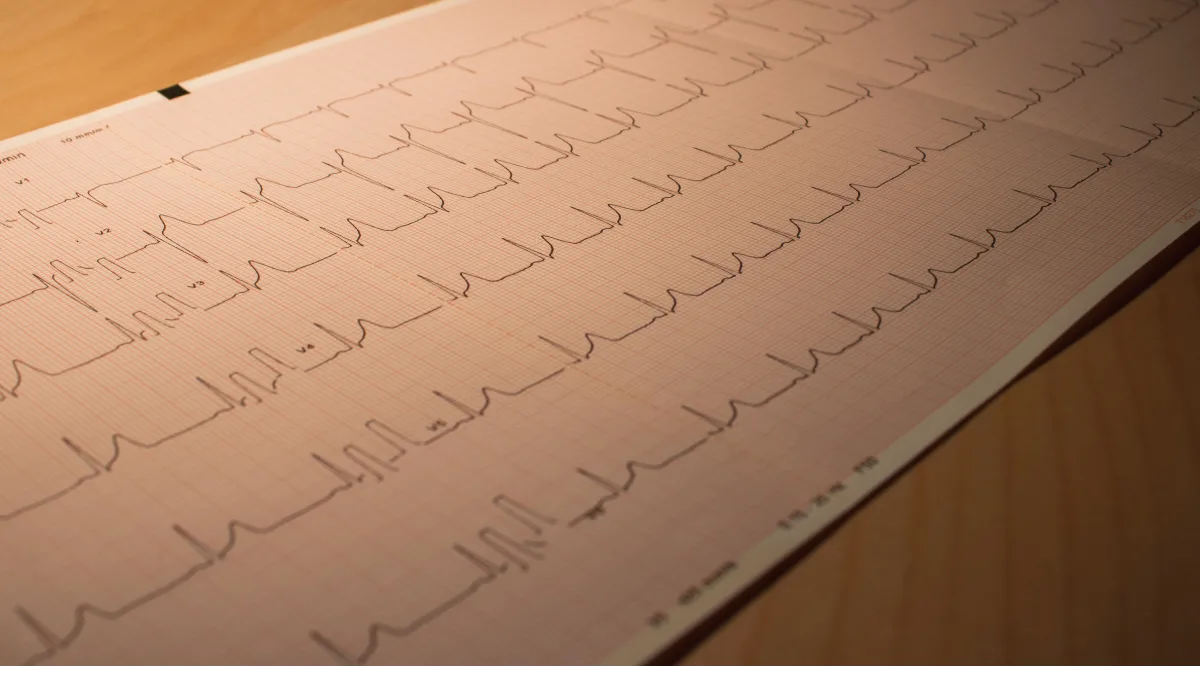
Because the electrodes are placed at different positions around the body, the ECG effectively looks at the heart’s electrical activity from 12 different angles, called leads or viewpoints. Each produces its own waveform on the paper trace, and together they give a detailed picture of how electrical signals are moving through the heart.

What a Q wave is

Each heartbeat produces a characteristic shape on the ECG trace, a series of peaks and dips. A Q wave is simply a small downward dip at the beginning of the main electrical spike of each beat. In many of the 12 viewpoints, small Q waves are entirely normal, they reflect the normal direction in which the electrical signal travels through the heart at the start of each beat.

In certain viewpoints, particularly one called lead III, which looks at the heart from a specific angle determined by the position of the left arm and left leg electrodes, a Q wave is especially common as a normal finding. It can appear and disappear simply with a change in body position or a deep breath. It is not a sign of damage. It is a reflection of the angle at which that particular viewpoint happens to be looking at the heart.

What Would a Genuine Concern Actually Look Like?

When a heart attack damages an area of heart muscle permanently, that area becomes electrically silent, it no longer generates the normal electrical signals. The ECG viewpoints looking directly at that damaged area will show an abnormal Q wave as a result, deeper, broader, and more prominent than a normal variant Q wave.

But, and this is the critical point, a heart attack affecting any meaningful area of muscle will show these changes across multiple neighbouring ECG viewpoints simultaneously, not in just one. It will also typically be accompanied by other supporting changes in the same viewpoints, changes in the shape of the waveform, and changes in the pattern of recovery between beats.

An isolated Q wave appearing in just one viewpoint, with every other viewpoint completely normal and no supporting changes anywhere on the trace, does not fit this picture at all. It is simply not how genuine heart attack scarring presents on an ECG. The same principle applies to other findings that sound alarming in isolation, such as a first-degree AV block, where context determines everything.

Why the Software Gets It Wrong

Modern ECG machines include automated interpretation software that analyses the trace and generates a written report. This software is useful, it can reliably identify certain patterns and flag them for clinical review. But it has an important limitation: it cannot think clinically.

When the software sees a Q wave in a particular viewpoint, it flags “possible old heart attack, clinical correlation recommended.” It cannot consider that the person is 32 years old and plays sport twice a week. It cannot consider that the Q wave is tiny and only visible in one viewpoint. It cannot consider that the person has never had any cardiac symptom in their life. It simply matches the pattern and generates the flag.

That flag is not a diagnosis. It is a prompt for a clinician to look at the full picture, and when a clinician does, the picture is almost always entirely reassuring.

In most of these referrals, one look at the ECG in the context of the patient in front of me resolves the question immediately. The report did its job. The clinical review does the rest.

Prof. Peter Barlis, Interventional Cardiologist

Normal Variant vs Genuine Concern, Plain Language Guide

Feature Almost certainly a normal variant Worth investigating further
How many viewpoints show the Q wave Only one viewpoint on the entire ECG Multiple neighbouring viewpoints showing the same change
The rest of the ECG Completely normal in every other respect Other changes present in the same viewpoints
The person’s history No cardiac symptoms ever, no risk factors, fit and active History of chest pain, breathlessness, or cardiovascular risk factors
Does it change with breathing Q wave reduces or disappears with a deep breath Persistent regardless of position or breathing
Why the ECG was done Routine, pre-employment, or incidental finding ECG done because of symptoms or known cardiac history
Echocardiogram result Normal heart structure and function throughout Abnormal muscle movement in the area the Q wave viewpoints correspond to

What Investigation Is Actually Needed?

A clinical review, not a cascade of tests

The appropriate response to a report like this in an otherwise healthy person is a clinical review with a cardiologist, not an immediate referral for a stress test, a CT scan of the coronary arteries, or a coronary angiogram. A cardiologist looking at the trace alongside your history and examination can in most cases answer the question definitively without any further testing at all.

If any uncertainty remains after that review, perhaps because there are some cardiovascular risk factors present, or because the ECG changes are borderline, an echocardiogram is the most efficient next step. This is an ultrasound of the heart that shows how the heart muscle is moving. If the muscle in the area corresponding to the Q wave viewpoint is moving completely normally, which it almost always is in these situations, that is powerful additional reassurance that no significant heart attack has occurred.

What you do not need

A fit, active, symptom-free person with no cardiovascular risk factors whose ECG shows an isolated Q wave in a single viewpoint, with everything else normal, does not need urgent investigation. There is no reason to stop exercising while waiting for a result. What is needed is a clinical review that puts the automated report in its proper context, and in most cases that conversation is the only investigation required.

What Is Worth Holding Onto

An automated ECG report is generated by software, not by a cardiologist. Its job is to flag things for clinical review, not to make diagnoses, and it has no way of knowing anything about the person attached to the trace.

A Q wave appearing in just one viewpoint, with everything else completely normal, is almost always a normal finding in a healthy heart. The pattern that genuinely indicates a previous heart attack appears across multiple neighbouring viewpoints at once, never in isolation.

A cardiologist reviewing the ECG alongside your history will in most cases give a clear and reassuring answer without further testing. Where any doubt remains, an echocardiogram provides the most direct additional reassurance.

Heart Matters Resource

When in Doubt, Get Checked Out

Where an ECG report mentions Q waves or a possible old heart attack, a cardiology review normally answers the question efficiently and very reassuringly. That anxiety is worth resolving properly rather than carrying it.

Read: When in Doubt, Get Checked Out →

Conclusion

The automated ECG report that says “possible old heart attack” is one of the most anxiety-generating phrases in cardiology, and in a fit, healthy, symptom-free person it is almost always an over-call by software that cannot apply clinical judgement. The Q wave it has flagged is real. The interpretation it has placed on that finding is almost certainly wrong in this context.

A Q wave appearing in just one ECG viewpoint, with no other changes anywhere on the trace, in a person who has never had cardiac symptoms and has no significant risk factors, is a normal finding. It does not mean your heart is damaged. It does not mean you have had a heart attack. And it does not mean you need urgent investigation.

What it means is that you need a cardiologist to look at your ECG and your history together, and give you the reassurance that the software, by its nature, simply cannot provide.

Related Reading

A Living Legend of Interventional Cardiology: Professor Patrick Serruys

heartmatters.com 2026 04 06T204658.338 1
Key Points

  • Professor Patrick Serruys is one of the most influential figures in the history of interventional cardiology, with over 3,500 peer-reviewed publications and 250,000 citations.
  • He introduced balloon angioplasty to the Netherlands in 1980 and performed the country’s first coronary stent implantation in 1986.
  • He helped pioneer drug-eluting stents, now the global standard of care, implanted in millions of patients every year.
  • In 2004, he performed the first percutaneous aortic valve replacement in the Netherlands, a procedure now known as TAVI.
  • He remains scientifically active today, continuing to shape the future of cardiovascular medicine.

Last week, I had the honour of presenting a lifetime achievement award to a man who shaped not only my career, but the entire field of interventional cardiology. Professor Patrick Serruys visited Sydney, and standing in front of him with that award in my hands, I found myself thinking about the extraordinary distance modern heart medicine has travelled, and how much of that journey he personally led.

Patrick was my PhD supervisor. He wrote the foreword to my book on heart stents. We continue to collaborate to this day. But this article is not really about my connection to him, it is about what his work means for patients. Because if you or someone you love has ever had a coronary stent, a drug-eluting stent, or a catheter-based heart valve procedure, there is a very real chance that the treatment you received exists in its current form because of Professor Serruys.

Where It All Began

Patrick Serruys published his first scientific paper in the British Heart Journal in 1978. He was working at the Thoraxcenter in Rotterdam, then a young institution that would become one of the most important centres of cardiovascular innovation in the world. From the very beginning, he was drawn to a question that would define his career: could blocked heart arteries be treated without open-heart surgery?

At the time, the answer was far from obvious. Coronary artery bypass surgery was the standard of care. The idea that a cardiologist could thread a thin catheter through the blood vessels, navigate to a blocked artery in the heart, and open it from the inside, without a single incision on the chest, was genuinely radical.

In September 1980, Professor Serruys introduced balloon angioplasty to Rotterdam. A small balloon on the tip of a catheter, inflated inside the narrowed artery to compress the blockage and restore blood flow. It worked. But it had a significant problem, the artery often narrowed again within months, a process called restenosis. For more than a decade, he led thirteen clinical trials attempting to solve this problem with medications. The results were disappointing.

The history of medicine is full of researchers who, faced with repeated setbacks, simply kept going. What distinguishes Professor Serruys is that each disappointment redirected his curiosity rather than diminishing it. The solution to restenosis, it turned out, was not a drug, it was a device.

The Stent That Changed Everything

In 1986, Professor Serruys performed the first coronary stent implantation in the Netherlands, just months after the very first procedures anywhere in the world. A coronary stent is a tiny mesh scaffold, deployed inside the artery to hold it open after balloon angioplasty. It was a transformative development. Restenosis rates fell. Patients did better.

But the stent itself still caused some degree of restenosis in a proportion of patients, because the metal triggered a healing response from the artery wall that could cause re-narrowing over time. The next challenge was clear: could the stent itself deliver medication directly to the artery wall to prevent this response?

By the late 1990s, working with colleagues in Rotterdam and São Paulo, Professor Serruys helped pioneer the first drug-eluting stents stents coated with medication that releases slowly into the surrounding tissue, dramatically reducing restenosis. In 2000, during one of cardiology’s most prestigious lectures, he predicted this technology would spread worldwide. It did. Drug-eluting stents are now the global standard of care for coronary intervention, implanted in millions of patients every year.

In 1994, he led the first randomised controlled trial directly comparing stenting with balloon angioplasty alone, published in the New England Journal of Medicine, which contributed to regulatory approval of coronary stents by the United States FDA that same year. If you want to understand the evidence behind the stent in your own chest, you can read more on our Coronary Artery Disease page.

Beyond the Stent

Even as stenting transformed interventional cardiology, Professor Serruys was already thinking about its limitations. A permanent metallic scaffold left forever inside a coronary artery troubled him. What if the scaffold could dissolve once its job was done, leaving the artery free and natural?

In 2006, he introduced fully biodegradable coronary scaffolds made from polylactic acid, the same material used in dissolvable surgical sutures, that provided the structural support of a stent during the critical healing period, then gradually disappeared over two to three years. The concept and early results were published in The Lancet and the New England Journal of Medicine. This remains an active and evolving area of research.

His curiosity never stayed confined to coronary arteries. In 2004, together with the pioneering French cardiologist Alain Cribier, he performed the first percutaneous aortic valve replacement in the Netherlands threading an artificial heart valve through the blood vessels and implanting it inside the diseased native valve, without open-heart surgery. This procedure, now known as TAVI, has since transformed the treatment of aortic stenosis and is now offered to tens of thousands of patients worldwide who previously had no good surgical option.

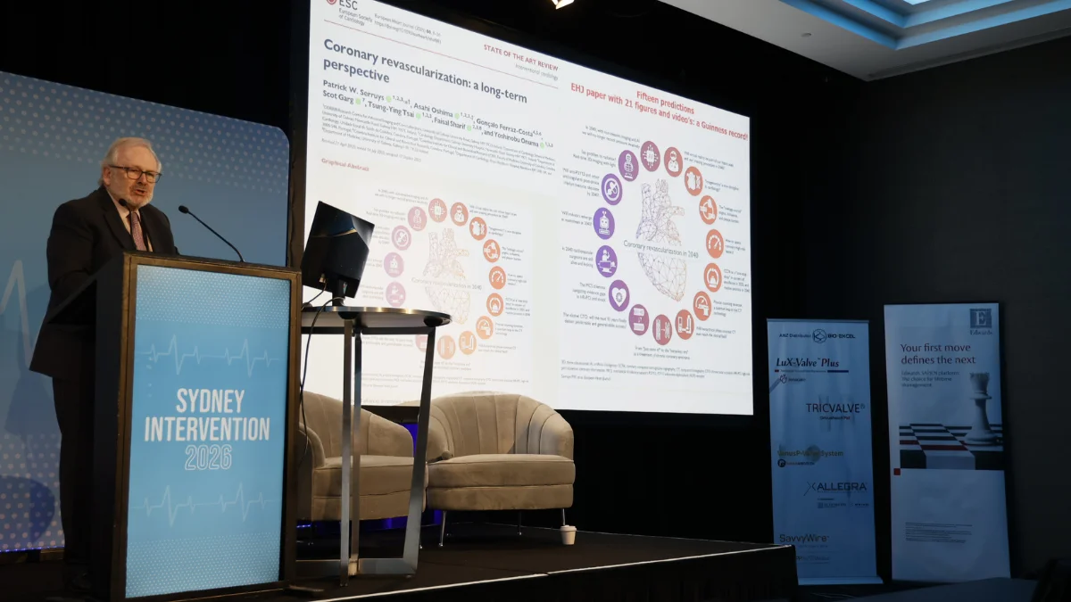
Professor Patrick Serruys presenting at Sydney Intervention 2026
Professor Serruys presenting at Sydney Intervention 2026, his lecture on the future of coronary revascularisation included fifteen predictions for the field, published in the European Heart Journal.

The Scale of a Career

Numbers can feel abstract, but in this case they help convey something genuinely difficult to communicate in words. Professor Serruys has published more than 3,500 peer-reviewed scientific papers. His work has been cited more than 250,000 times by other researchers, placing him among the most cited medical scientists on the planet.

He has trained more than 400 interventional cardiologists and supervised more than 100 PhD candidates many of whom are now leading figures in the field in their own right. I am proud to count myself among them.

He is the author or co-author of 43 books and monographs, including three editions of the European Society of Cardiology’s flagship textbook of cardiovascular medicine. He holds an honorary doctorate in engineering from the University of Melbourne, a recognition that his contributions straddled the boundary between clinical medicine and biomedical engineering.

At the time of writing, he remains scientifically active at the University of Galway, where he established a research centre focused on advanced imaging and core laboratory science after his 36-year career at Erasmus University in Rotterdam. He cycles to the laboratory every day.

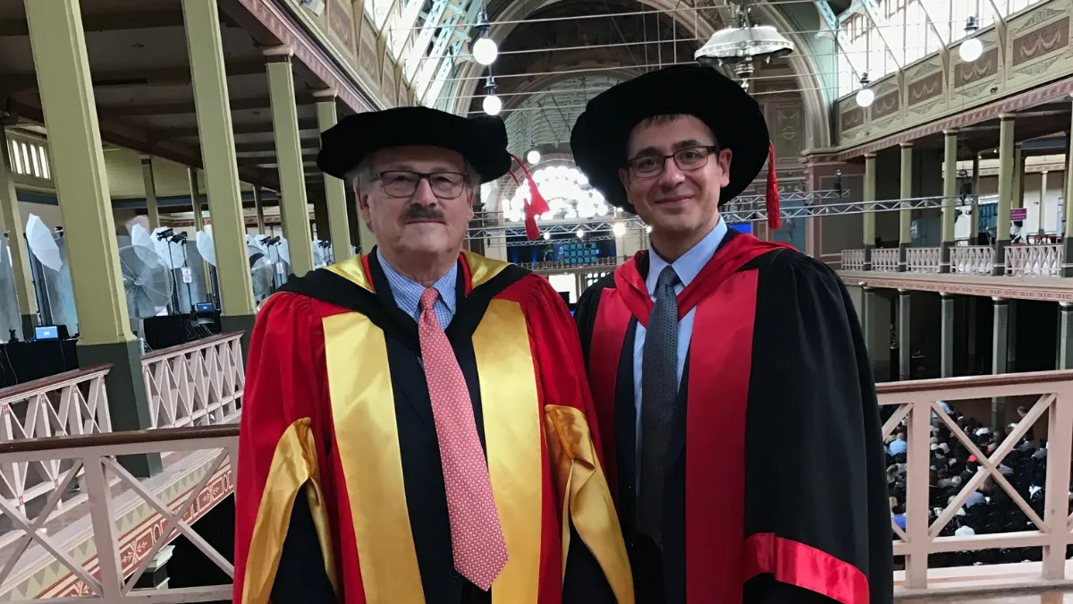
Professor Patrick Serruys and Prof. Peter Barlis at the University of Melbourne honorary doctorate ceremony 2016
Professor Serruys receiving his honorary doctorate in engineering from the University of Melbourne in 2016, pictured with Prof. Peter Barlis.

What This Means for Patients

I am sometimes asked by patients why any of this history matters to them. The answer is simple. Every time a cardiologist threads a stent into a blocked coronary artery, a procedure that takes less than an hour, requires no general anaesthetic, and sends most patients home the same day, they are building on decades of work by researchers like Professor Serruys who refused to accept that open-heart surgery was the only answer.

The treatments we now consider routine were once considered impossible. They exist because of people who asked difficult questions, ran rigorous trials, published honest results, including failures, and kept pushing. Understanding that journey helps patients engage more confidently with their own care.


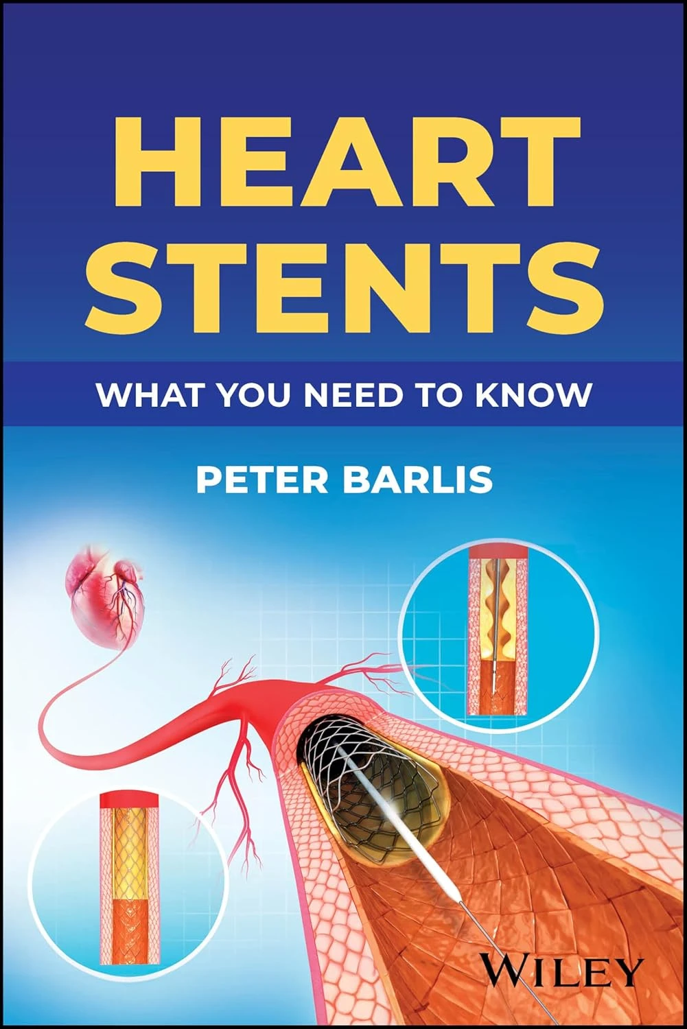
Heart Stents: What You Need to Know by Prof. Peter Barlis

New Release 2026

Heart Stents: What You Need to Know

A comprehensive guide by Professor Peter Barlis, with a foreword by Professor Patrick Serruys. Published by Wiley.


Buy on Amazon →

Conclusion

Presenting that lifetime achievement award to Patrick last week, in Sydney, surrounded by colleagues whose careers he has shaped, was one of the genuine privileges of my professional life. The field of interventional cardiology owes him an enormous debt.

And so, indirectly, do the millions of patients whose lives have been changed by the treatments he helped bring into existence. If you have ever had a stent placed, a valve replaced without open-heart surgery, or benefited from any of the imaging technologies now used in the catheterisation laboratory, there is a very good chance that Professor Serruys played a role in making that possible.

That is a legacy worth celebrating, not just within cardiology, but for every patient who has sat in a recovery room, gone home the next morning, and returned to their life.

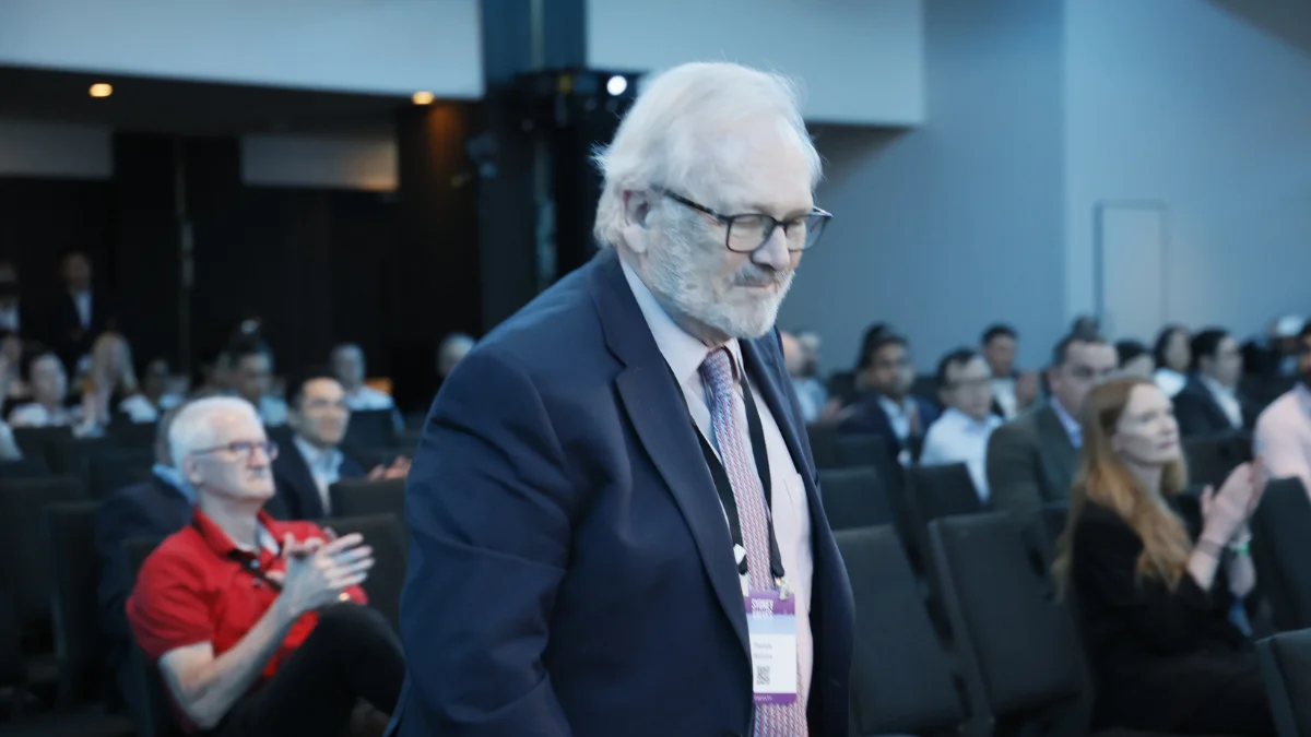
Professor Patrick Serruys receives a standing ovation at Sydney Intervention 2026
Sydney Intervention 2026, a room full of cardiologists rises as Professor Serruys receives the lifetime achievement award.

More from Heart Matters: Coronary Artery Disease · Heart Stents Explained · TAVI, What to Expect · When in Doubt, Get Checked Out

Cold Hands and Feet: Causes, Circulation and What to Watch For

cold hands
Key Points

  • Cold hands and feet are very common, and in most cases they reflect normal thermoregulation rather than disease. For many people the problem is transient or weather-related and signals nothing of concern.
  • When cold hands and feet come and go with the temperature, your mood, or the season, that pattern is reassuring. It is persistent, one-sided, or progressive coldness that is worth a closer look.
  • If the problem is ongoing, the causes worth considering widen to include peripheral arterial disease, Raynaud’s phenomenon, an underactive thyroid, anaemia, diabetes, and certain medications, particularly beta-blockers.
  • Peripheral arterial disease (PAD), narrowing of the arteries that supply the legs, can cause persistently cold feet, especially alongside leg pain on walking, skin colour changes, or slow-healing wounds.
  • A small number of straightforward tests, a pulse check, an ankle-brachial pressure index, and a few blood tests, can usually sort the benign from the significant. Ongoing or worsening symptoms are worth discussing with your doctor.

Cold hands and feet are one of the most commonly reported symptoms in general practice, and one of the most frequently dismissed. “Poor circulation” is the phrase most patients are given, which is vague enough to cover everything from completely normal thermoregulation to significant arterial disease.

Here is the reassuring part, and it applies to most people reading this. Cold hands and feet are very often transient. They track the weather, the season, a cold office, a drop in activity, or even stress and tiredness. When that is the pattern, the coldness is the body behaving exactly as it should, and it points to nothing wrong with the heart or the circulation at all.

For some people, though, cold extremities are the first sign of a condition that shares the same underlying process as coronary artery disease, and deserves the same attention. The practical value of this article is knowing which pattern you are dealing with, and what to do if the coldness is ongoing rather than coming and going.

Why Extremities Feel Cold

Normal thermoregulation

The body’s first priority in cool conditions is maintaining its core temperature. The nervous system does this by narrowing the blood vessels in the skin and extremities, reducing blood flow to the periphery so that warm blood keeps circulating to the brain, heart, and other vital organs. This is a normal, healthy response, and the hands and feet are the first places to feel it because they are furthest from the core.

Some people are more prone to it than others: women more than men, those with a slimmer build, and people with naturally lower blood pressure. In these individuals, cold hands and feet are a constitutional feature, not a disease. They tend to be lifelong, symmetrical, and closely tied to the temperature around them.

When it is reassuring, and when it is worth a closer look

The pattern usually tells the story. Coldness that comes and goes with the weather, eases when you warm up, affects both sides equally, and has been with you for years is reassuring. It does not need investigation.

The picture changes when cold extremities are persistent rather than intermittent, are clearly worse on one side, are getting steadily worse over months, or arrive alongside other symptoms such as leg pain on walking, skin changes, or wounds that will not heal. That is the shift from normal thermoregulation to something worth exploring, and the rest of this article walks through the possibilities.

Peripheral Arterial Disease

What it is

Peripheral arterial disease is atherosclerosis, the same process of gradual plaque build-up that affects the coronary arteries, occurring instead in the arteries that supply the legs and feet. As these narrowings progress over time, blood flow to the lower limbs becomes increasingly limited.

PAD and coronary artery disease share the same risk factors: smoking, diabetes, high blood pressure, high cholesterol, and a family history. The two conditions often coexist, which is why identifying PAD matters well beyond the legs themselves.

1 in 5
people over 65 have peripheral arterial disease, and most are unaware of it, often putting their symptoms down to ageing or general unfitness.
American Heart Association

Symptoms to be aware of

Cold feet in PAD usually come with other features that help separate it from ordinary cold sensitivity. Leg pain, cramping, or heaviness that comes on with walking and eases with rest, known as intermittent claudication, is a common feature of significant PAD. More advanced disease can cause pain in the foot or toes at night. Skin changes are also worth noting: pallor, shiny skin, loss of hair on the feet and lower legs, or wounds that are slow to heal. Any of these are worth bringing to your doctor’s attention.

Reduced or absent foot pulses in someone with cold feet, leg pain on walking, and a history of smoking or diabetes is worth investigating. It changes the cardiovascular risk conversation in an important way.

Prof. Peter Barlis, Interventional Cardiologist

Raynaud’s Phenomenon

Raynaud’s phenomenon is a different and very common condition. It is not fixed narrowing in the arteries but brief episodes of arterial spasm triggered by cold or emotional stress. The classic pattern is a sequence of colour changes in the fingers: they turn white as blood flow briefly stops, then blue as blood pools, then bright red as circulation returns. The episode is usually accompanied by numbness and tingling, and can feel uncomfortable as the fingers rewarm.

The reassuring news is that primary Raynaud’s, which occurs on its own without any underlying disease, is extremely common, more frequent in women, and in the large majority of cases entirely benign. Keeping the hands warm, avoiding sudden cold exposure, and managing stress are the main practical measures.

A less common form, secondary Raynaud’s, can occasionally be linked to connective tissue conditions. Features that might prompt your doctor to look further include a later age of onset, symptoms affecting only one hand, or associated joint pain or skin changes. If you have Raynaud’s and any of these apply, it is worth mentioning at your next appointment.

Other Causes Worth Considering

When cold hands and feet are ongoing rather than weather-related, it is worth remembering that the cause is often not the arteries at all. Several common, treatable conditions reduce peripheral warmth, and most are picked up with a simple blood test.

An underactive thyroid slows the whole metabolism and is one of the most frequent reasons for feeling cold all over, the hands and feet included, often with tiredness, weight gain, and dry skin alongside. Anaemia, where there are too few red cells to carry oxygen efficiently, can leave the extremities cold and pale and is common in women of menstruating age. Diabetes contributes in two ways, by affecting the small blood vessels and by causing nerve changes in the feet that alter how temperature is sensed. Smoking narrows blood vessels directly and is one of the most powerful and modifiable contributors of all. A naturally low body weight, low blood pressure, dehydration, and even anxiety, which diverts blood away from the skin, can all play a part.

None of these is a reason for alarm. They are listed here because they are common, they are easily tested for, and they respond well to treatment once identified.

Medications and the Heart

Sometimes the cause is not the arteries or the metabolism, but a medication. This is worth knowing, because it is common, often reversible, and easy to miss.

Beta-blockers, used widely in cardiovascular medicine for conditions including high blood pressure, heart failure, and recovery after a heart attack, can reduce blood flow to the hands and feet and make cold extremities noticeably worse. This is a known and recognised side effect, and it tends to be most noticeable in people who already have Raynaud’s or cold sensitivity. If your cold hands and feet started or became much worse after you began a beta-blocker, it is worth raising with your prescribing doctor. There are often alternatives, and your team will weigh the benefit of the medication against this side effect with you.

Reduced pumping power of the heart is a separate and more significant cause. In heart failure, the heart cannot maintain its normal output, and the body responds by diverting blood away from the skin and extremities to protect the vital organs. The result can be cold, pale, sometimes mottled hands and feet, usually as part of a broader picture that includes breathlessness, fatigue, and swelling of the ankles. Cold extremities on their own are not a sign of heart failure, but cold extremities alongside those other symptoms are worth prompt assessment.

Feature Normal / Raynaud’s Peripheral Arterial Disease
Distribution Both hands and feet symmetrically Often worse in feet; may be asymmetric
Colour changes White, blue, then red sequence (Raynaud’s) Pallor or dusky discolouration, no rewarming flush
Pain with walking Not typically Leg cramping or heaviness with exertion
Skin changes Normal skin Shiny skin, hair loss, slow wound healing
Pulses Normal Reduced or absent foot and ankle pulses

What Helps Day to Day

For the large majority of people, whose cold hands and feet are constitutional or weather-related, a few simple habits make a real difference. These measures are also the sensible first step while you arrange to have any ongoing symptoms checked.

Warm the core, not just the hands

The body warms the extremities last. A warm torso, a hat, and layered clothing do more for cold fingers and toes than gloves alone.

Keep moving

Regular activity improves circulation throughout the body. Even a short walk or wiggling the toes and fingers brings warmth back to cold extremities.

Avoid the triggers

Not smoking is the single most important step for circulation. Limiting caffeine and managing stress also help, as both can narrow small blood vessels.

When to See Your Doctor and What Tests to Expect

Cold hands and feet without any worrying features, in someone with no cardiovascular risk factors, normal skin, no leg pain on walking, and a lifelong weather-related pattern, generally do not need investigation. The day-to-day measures above are enough.

It is worth seeing your doctor when the coldness is persistent rather than intermittent, affects one side more than the other, is getting worse over time, or comes with leg pain on walking, skin changes, wounds that are slow to heal, or significant cardiovascular risk factors such as smoking, diabetes, or high blood pressure. The assessment is straightforward and usually involves no more than the following.

Your doctor will start by feeling the pulses in your feet and ankles, which gives an immediate sense of how well blood is reaching the lower limbs, and by examining the skin. An ankle-brachial pressure index, a painless test that compares the blood pressure reading at the ankle with the reading at the arm, gives a reliable picture of arterial flow to the legs and can be done at a routine appointment. A few blood tests are commonly arranged at the same time to check the thyroid, look for anaemia, and assess blood glucose. If Raynaud’s appears to be secondary rather than primary, your doctor may add an autoimmune blood screen or look at the small vessels at the base of the fingernails. Where PAD is suspected, an ultrasound scan of the leg arteries can map exactly where any narrowing lies.

Questions Worth Asking at Your Next Appointment

  • My cold hands and feet are ongoing rather than weather-related. Could they reflect reduced blood flow rather than normal cold sensitivity?
  • Should I have my pulses checked and an ankle-brachial pressure index done?
  • Could a thyroid problem, anaemia, or my blood sugar be contributing? Are those worth testing?
  • My beta-blocker seems to have made my cold hands worse, is there an alternative worth considering?
  • I have Raynaud’s, is there anything that suggests it could be related to an underlying condition?
  • I have a wound on my foot that is not healing as expected, when should I have this assessed?

Heart Matters Resource

When in Doubt, Get Checked Out

Cold feet with leg pain on walking, skin changes, or a wound that is not healing as expected is worth a conversation with your doctor. Peripheral arterial disease is treatable when it is found early, and early assessment makes a real difference.

Read: When in Doubt, Get Checked Out

Conclusion

Cold hands and feet are usually benign, the body doing exactly what it is designed to do, putting core temperature ahead of peripheral comfort. For most people the explanation is constitutional or weather-related, the symptoms come and go, and no investigation is needed.

When the coldness is ongoing, one-sided, or worsening, or comes with leg pain, skin changes, or other symptoms, the list of causes widens to include peripheral arterial disease, an underactive thyroid, anaemia, diabetes, and medication effects, all of which are readily tested for and treatable. The line between cold feet that are normal and cold feet worth discussing is usually clear from the pattern. If you are not sure which side you fall on, that uncertainty is itself a good reason to ask.

Related Reading

Obstructive Sleep Apnoea and the Heart: Why It’s So Often Missed

obstructive sleep apnoea
Key Points

  • Obstructive sleep apnoea, often called OSA or simply sleep apnoea, is a condition where the airway repeatedly closes during sleep, causing breathing to stop briefly, sometimes hundreds of times a night. Most people have no idea it is happening.
  • Sleep apnoea is common. Around one in ten adults has the moderate-to-severe form with the clearest cardiovascular consequences, and rates are even higher when milder cases are included. The vast majority of people who have it have never been diagnosed.
  • The heart connections are significant and well established. Sleep apnoea is independently linked to high blood pressure, atrial fibrillation, heart failure, and increased risk of heart attack and stroke.
  • Symptoms include loud snoring, waking feeling exhausted despite adequate sleep, morning headaches, and significant daytime tiredness. Many people have subtle symptoms and do not recognise the pattern.
  • Treatment with CPAP, a small device that keeps the airway open during sleep, is highly effective and produces real cardiovascular benefits including better blood pressure control and reduced atrial fibrillation burden.

Most people who have sleep apnoea do not know they have it. They know they snore. They know they feel tired no matter how much they sleep. They know they wake with a headache some mornings. But they have put these things down to age, to stress, to being overweight, and the possibility that something specific and very treatable is happening during the night has never been raised.

Sleep apnoea sits at the junction of sleep health and heart health in a way that medicine has taken too long to fully recognise. It is not simply a snoring problem. It is a condition that stresses the cardiovascular system repeatedly through the night, and when it is identified and treated, the benefits extend well beyond simply sleeping better.

This article is for anyone who recognises the pattern described here, and for anyone managing a heart condition who has never been assessed for a sleep disorder that may be quietly working against their treatment.

What Is Sleep Apnoea?

What happens during the night

During sleep, the muscles throughout the body relax, including the muscles that support the soft tissues of the throat. In most people this relaxation is harmless. In people with obstructive sleep apnoea, the airway partially or completely collapses when those muscles relax, blocking the flow of air to the lungs.

When breathing stops, the level of oxygen in the blood begins to fall. The brain detects this and briefly rouses the person, just enough to restore muscle tone in the throat and allow breathing to restart, usually with a snort, gasp, or choking sound. The person rarely fully wakes and rarely remembers any of this. But it may happen dozens or even hundreds of times through the night, preventing the deep, restorative stages of sleep and leaving the person exhausted in the morning despite apparently adequate hours in bed.

How severity is measured

Sleep specialists measure the severity of sleep apnoea by counting the average number of breathing interruptions per hour of sleep. Mild sleep apnoea involves 5 to 15 events per hour. Moderate is 15 to 30. Severe is above 30, which means breathing is being interrupted more than once every two minutes throughout the night. In some people with untreated severe sleep apnoea, this happens every single minute of sleep, a level of overnight stress on the body that has very real cardiovascular consequences.

Recognising the Symptoms

Sleep apnoea has a recognisable pattern, but many of its features are easy to dismiss as something else. The symptoms below are the most useful to identify.

Symptoms to Recognise

Loud Snoring

Often loud enough to wake a partner. Not every snorer has sleep apnoea, but significant sleep apnoea is almost always accompanied by snoring.

Witnessed Pauses

A partner notices the person stop breathing and restart with a snort or gasp. If someone has told you this, it is one of the most important things to mention to your doctor.

Unrefreshing Sleep

Waking after a full night still feeling exhausted. Many people normalise this over years without realising it reflects a treatable condition.

Morning Headache

A dull pressure headache on waking that clears within an hour of getting up. Caused by overnight build-up of carbon dioxide in the blood.

Daytime Sleepiness

Struggling to stay awake during meetings, in front of the television, or while reading. Falling asleep at the wheel is a real risk in significant untreated sleep apnoea.

Nocturnal Palpitations

Each breathing pause triggers a stress surge that can disturb heart rhythm overnight, particularly relevant in atrial fibrillation.

If morning headaches or palpitations at night are a regular feature for you, our articles on morning headaches and the heart and why you feel your heart beating at night explore both connections in more detail.

Who Is at Risk?

Sleep apnoea is more common in men than women, though the gap narrows significantly after menopause. Carrying extra weight, particularly around the neck, is the most important modifiable risk factor, as it narrows the airway. Getting older, having a naturally narrow jaw, large tonsils, or a blocked nose all increase the likelihood of sleep apnoea developing.

But sleep apnoea is not exclusively a condition of overweight middle-aged men, and this assumption causes many people to go undiagnosed. Lean individuals, women, and younger people all develop sleep apnoea. People of East and Southeast Asian background develop it at lower body weights than Western populations due to differences in facial bone structure. The symptoms matter more than the stereotype. If the pattern fits, it is worth raising with your doctor regardless of what you weigh or what age you are.

Why Sleep Apnoea Matters for Your Heart

High blood pressure

Sleep apnoea is the most common identifiable cause of blood pressure that is difficult to control, where pressure stays high despite medication. The reason is that each time breathing stops during the night, the body’s stress response fires, raising the heart rate and tightening the blood vessels. This happens so many times through the night that the stress response carries over into the daytime, keeping blood pressure elevated around the clock.

For anyone whose blood pressure has been hard to bring under control despite treatment, asking about sleep apnoea is one of the most valuable steps available. Treating the sleep apnoea often produces blood pressure improvements that medication alone could not achieve.

Atrial fibrillation

Atrial fibrillation, an irregular heart rhythm that significantly increases stroke risk, is closely connected to sleep apnoea. The two conditions frequently coexist, and untreated sleep apnoea makes atrial fibrillation harder to treat and more likely to return after treatment. The overnight oxygen drops and stress surges from sleep apnoea irritate the heart’s electrical system in ways that promote irregular rhythm.

For anyone who has had cardioversion, an electrical reset of the heart rhythm, or catheter ablation to treat AF, treating sleep apnoea is now considered a standard part of protecting that result. Without it, the AF is significantly more likely to return.

Heart failure

In people with heart failure, where the heart is not pumping as efficiently as it should, sleep apnoea adds an additional burden on the heart through the night, at the very time the heart should be resting and recovering. Treating sleep apnoea in people with heart failure improves the heart’s pumping function and reduces the overnight stress load.

Heart attack and stroke risk

The repeated overnight stress that untreated sleep apnoea places on the blood vessels accelerates the build-up of plaque in the arteries, the same process that underlies heart attacks and strokes. Sleep apnoea is an independent cardiovascular risk factor, meaning it adds to risk over and above the conventional factors like blood pressure, cholesterol, and smoking.

Getting a Diagnosis: Simpler Than You Might Think

The sleep study

Diagnosing sleep apnoea requires a sleep study, but this is far simpler than most people imagine. The most common approach is a home-based study, a small portable monitor worn overnight in your own bed. It measures oxygen levels, breathing patterns, heart rate, and body position through the night. Most people sleep almost normally wearing it. The results are reviewed by a sleep specialist and used to determine whether sleep apnoea is present and how severe it is.

A formal in-laboratory sleep study, where the person sleeps overnight at a clinic with more detailed monitoring, is sometimes used for more complex cases, but the home study is the standard starting point for most people.

How to access a sleep study

Your GP or cardiologist can arrange a referral for a sleep study. If you have established cardiovascular disease, particularly high blood pressure that is hard to control, atrial fibrillation, or heart failure, and sleep apnoea has never been assessed, raising it proactively at your next appointment is worthwhile. Many people have been managing their heart condition for years without this important piece of the picture being investigated.

Treatment: What Works and What to Expect

CPAP: The Most Effective Treatment

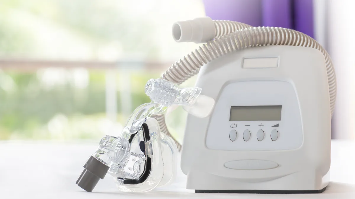
CPAP, which stands for Continuous Positive Airway Pressure, is the most effective treatment for moderate to severe sleep apnoea. It involves wearing a mask during sleep that delivers a gentle, steady flow of air. This air pressure acts like a splint, keeping the airway open and preventing it from collapsing throughout the night.

Modern CPAP machines are much quieter and more comfortable than earlier generations. Many people are surprised by how unobtrusive they are in practice. The mask comes in several styles, and finding the right fit makes a significant difference to comfort. Most people go through an adjustment period of two to four weeks, and the vast majority who persist through that period find the improvement in their sleep, their daytime energy, and their overall wellbeing to be genuinely transformative.

The cardiovascular benefits of consistent CPAP use are real and measurable: better blood pressure control, reduced atrial fibrillation burden, improved heart function in heart failure, and lower overnight cardiovascular stress. For many people, CPAP treatment changes not just their sleep but their overall cardiac management picture.

Weight loss

For people who are overweight, meaningful weight loss reduces the severity of sleep apnoea significantly, and in some cases resolves it entirely. This is the most durable long-term solution. In practice, CPAP and weight loss often work together. The CPAP provides immediate protection while lifestyle changes work over time.

Sleeping position and dental devices

For milder sleep apnoea, particularly in people whose apnoeas mainly occur when sleeping on their back, simply learning to sleep on the side can make a meaningful difference. Custom dental appliances that gently advance the lower jaw during sleep are another option for people with mild to moderate sleep apnoea who cannot tolerate CPAP. They are made by a dentist with experience in sleep disorders and can be very effective in the right patient.

Sleep apnoea assessment is now a routine part of how I evaluate patients with high blood pressure, AF, and heart failure. When it is present and treated, the difference to their cardiac management can be substantial. Treating the heart condition without addressing the sleep apnoea is working with one hand tied behind your back.

Professor Peter Barlis, Interventional Cardiologist

Questions Worth Raising with Your Doctor

  • I snore heavily and wake feeling exhausted regardless of how long I sleep. Should I be assessed for sleep apnoea?
  • My blood pressure has been difficult to control despite medication. Could untreated sleep apnoea be a factor?
  • I have atrial fibrillation. Has sleep apnoea been assessed as part of my management?
  • I have heart failure. Should a sleep study be part of my investigation?
  • I have started CPAP but am finding it difficult to get used to. What support is available?

Heart Matters Resource

When in Doubt, Get Checked Out

If you recognise the pattern described in this article, or if your partner has raised concerns about your breathing during sleep, a sleep study is a straightforward, low-barrier investigation that can answer the question definitively. Raise it with your GP or cardiologist at your next appointment.

Read: When in Doubt, Get Checked Out →

Conclusion

Sleep apnoea is common, under-diagnosed, and very treatable. The connection to cardiovascular health is real and significant, and identifying it in someone managing high blood pressure, atrial fibrillation, or heart failure can genuinely change their clinical picture for the better.

The home sleep study is simple, the treatment is effective, and the improvement in how people feel, in their sleep, their energy, their capacity to engage with life, is one of the most consistent and satisfying outcomes in all of cardiovascular medicine.

If any of the symptoms in this article sound familiar, that conversation with your doctor is worth having. A good night’s sleep is not a luxury. For your heart, it is part of the treatment plan.

Related Reading

POTS: Understanding Postural Orthostatic Tachycardia Syndrome

heartmatters.com 2026 03 31T224224.252
Key Points

  • POTS, Postural Orthostatic Tachycardia Syndrome, is a condition of the autonomic nervous system in which heart rate rises excessively on standing, producing symptoms that can be profoundly disabling.
  • The hallmark is a heart rate increase of 30 beats per minute or more within 10 minutes of standing, without a significant fall in blood pressure, accompanied by a characteristic symptom pattern.
  • Symptoms include dizziness, lightheadedness, palpitations, fatigue, brain fog, and near-fainting on standing, often dramatically improved by lying down, which is one of the most diagnostically telling features.
  • POTS is not rare, it predominantly affects women between the ages of 15 and 50, and is significantly under-diagnosed. Many patients spend years being told their symptoms are anxiety or deconditioning before receiving a correct diagnosis.
  • While POTS can be debilitating, a structured management approach, combining lifestyle strategies, physical reconditioning, and where needed medications, produces meaningful improvement in the majority of patients. Specialist clinic input is an important part of optimal care.

POTS is one of those conditions where the journey to diagnosis is often as difficult as the condition itself. Patients, most of them young, arrive in my clinic having seen multiple doctors, having been told their palpitations are anxiety, their fatigue is depression, their dizziness is nothing to worry about. Some have been told there is nothing wrong. Others have been given a diagnosis of chronic fatigue, fibromyalgia, or panic disorder, and while these may coexist, they are not POTS, and treating them alone leaves the underlying autonomic dysfunction unaddressed.

The relief that comes when POTS is finally named and explained, when a patient understands that their symptoms have a physiological basis that is measurable and treatable, is one of the more meaningful moments in a consultation. It does not make the condition less challenging. But it makes it navigable.

This article is for patients who suspect they may have POTS, who have recently been diagnosed, or who are trying to understand a condition that is often poorly explained. The message I want to convey from the outset is this: POTS is real, it is complex, it is frequently underestimated, and with the right approach, most people do meaningfully better.

What Is POTS?

The autonomic nervous system

The autonomic nervous system regulates the body’s automatic functions, heart rate, blood pressure, breathing, digestion, without conscious effort. When you stand up, it orchestrates an immediate response: blood vessels in the legs constrict to prevent blood pooling downward, and the heart rate adjusts to maintain adequate blood flow to the brain. In most people this happens seamlessly and invisibly.

In POTS, this orchestration is dysregulated. When standing, blood pools excessively in the lower body. The autonomic nervous system compensates with a disproportionate surge in heart rate, but this response is not fully effective, and the brain and upper body receive inadequate perfusion. The result is the characteristic symptom cluster of POTS: dizziness, palpitations, fatigue, and cognitive fog that appear on standing and improve dramatically on lying down.

The diagnostic criteria

The formal diagnostic criterion for POTS is a sustained heart rate increase of 30 beats per minute or more within 10 minutes of standing, or a heart rate exceeding 120 beats per minute on standing, in the absence of orthostatic hypotension (a significant fall in blood pressure on standing). In adolescents, the threshold is a rise of 40 beats per minute.

The key distinction from a simple faint or vasovagal episode is that in POTS, the blood pressure does not fall significantly, it is the heart rate that is the primary abnormality, compensating for inadequate venous return with a dramatic and sustained tachycardia.

What Does POTS Feel Like?

The upright-to-horizontal contrast

One of the most revealing features of POTS, both for diagnosis and for the patient’s own understanding, is how dramatically symptoms vary with position. Standing or sitting upright produces symptoms. Lying down relieves them, often within minutes. This positional dependence is so characteristic that many patients learn to structure their lives around it before they have any diagnosis, lying down after meals, avoiding prolonged standing, sitting rather than standing whenever possible.

When patients describe having to lie on the supermarket floor, or being unable to stand in the shower, or feeling well in bed but incapacitated within minutes of getting up, that history is POTS until proven otherwise.

The symptom cluster

Palpitations

Racing heart on standing, often the most alarming feature. The heart rate surge is real and measurable, not imagined.

Dizziness and lightheadedness

On standing, prolonged standing, or after meals. Reflects inadequate cerebral perfusion despite the compensatory tachycardia.

Profound fatigue

Not ordinary tiredness, a heavy, persistent exhaustion that does not resolve with rest and is worsened by upright activity.

Brain fog

Difficulty concentrating, slowed thinking, memory problems. Reflects reduced cerebral blood flow rather than a primary neurological disorder.

Near-fainting (presyncope)

The feeling of being about to faint, often without actually losing consciousness. Many patients faint eventually but presyncope is more common.

Other autonomic features

Nausea, sweating, temperature dysregulation, headache, and sleep disturbance are common, reflecting the broader autonomic nervous system dysfunction.

Who Gets POTS and Why?

Demographics

POTS predominantly affects women, around 80% of cases, typically between the ages of 15 and 50. The onset is often in adolescence or young adulthood. It is estimated to affect between one and three million people in the United States alone, making it considerably more common than many conditions that receive far greater clinical attention.

Triggers and associations

POTS can develop after a viral illness, a pattern that has been particularly well documented following COVID-19, where post-COVID POTS has been identified as one of the more prevalent long COVID manifestations. Other recognised triggers include significant physical deconditioning, pregnancy, surgery, trauma, and puberty. In some patients there is no identifiable trigger, the autonomic dysregulation appears to be constitutional.

Associated conditions include hypermobile Ehlers-Danlos syndrome, a connective tissue disorder characterised by joint hypermobility, which is found in a significant proportion of patients with POTS. Mast cell activation syndrome, autoimmune conditions, and small fibre neuropathy are also more common in the POTS population than in the general population. These associations are clinically important because they influence investigation and management.

Subtypes

POTS is not a single pathophysiological entity, several distinct subtypes have been described, each with different underlying mechanisms. Hypovolaemic POTS involves a reduced circulating blood volume. Neuropathic POTS involves partial autonomic denervation of the lower limb blood vessels. Hyperadrenergic POTS involves excessive sympathetic nervous system activity. Understanding the subtype, where this is possible, helps guide treatment selection. This is one of the reasons specialist clinic input is so valuable.

Diagnosis

The active stand test

The simplest diagnostic assessment is the active stand test, measuring heart rate and blood pressure after lying supine for 10 minutes, then at intervals over 10 minutes of standing. A sustained heart rate rise of 30 beats per minute or more (40 in adolescents), with symptoms, and without significant blood pressure fall, meets the diagnostic criteria.

Tilt table testing

For a more controlled assessment, or when the active stand test is inconclusive, a tilt table test is performed. The patient is strapped to a table that is tilted from horizontal to 70 degrees and held there for up to 45 minutes while heart rate and blood pressure are continuously monitored. This test is the gold standard for diagnosing POTS and other forms of orthostatic intolerance.

Further investigation

Blood tests assess for common associated conditions, thyroid function, anaemia, autoimmune markers, and plasma volume studies where available. A 24-hour Holter monitor documents the heart rate patterns throughout a normal day. Echocardiography confirms normal cardiac structure and function. Skin biopsy for small fibre neuropathy may be considered in specialist centres.

One of the most validating moments for a patient with POTS is seeing their own heart rate trace on a monitor, watching it jump from 70 to 130 beats per minute simply on standing. For someone who has been told for years that their symptoms are anxiety or deconditioning, seeing the objective evidence of what their body is doing is genuinely transformative. It changes the conversation from “is this real?” to “what are we going to do about it?”, and that is a much better conversation to be having.

— Prof. Peter Barlis, Interventional Cardiologist

Management

Why a specialist clinic matters

POTS is best managed by a multidisciplinary team with experience in autonomic disorders. In Australia, POTS clinics, typically combining cardiology, neurology, and physiotherapy, offer the comprehensive, coordinated approach that this condition requires. A cardiologist managing a patient with POTS in isolation can help, but the physiotherapy reconditioning programme, the dietary advice, the psychological support for coping with a chronic and often poorly understood condition, these are best provided by a team that has developed expertise in this specific patient group.

If you have been diagnosed with POTS and are not yet under the care of a specialist clinic, asking for a referral is a worthwhile conversation to have.

Non-pharmacological strategies, the foundation

The core of POTS management is non-pharmacological, and for many patients, these measures alone produce significant improvement.

Fluid and salt loading is fundamental. Increasing fluid intake to two to three litres of water per day and increasing dietary salt, in the absence of hypertension, expands circulating blood volume and reduces the degree of orthostatic pooling. Many patients notice improvement within days of implementing this consistently.

Compression garments, waist-high graduated compression stockings or abdominal binders, physically counteract blood pooling in the lower body on standing. They are unglamorous but effective, and most patients who use them consistently find them meaningful.

Physical reconditioning is one of the most impactful and most challenging elements of management. Deconditioning worsens POTS significantly, yet upright exercise is poorly tolerated in active POTS. The key is starting with recumbent exercise, rowing machines, recumbent cycling, swimming, that achieves cardiovascular conditioning without the orthostatic stress of being upright. Gradually, as tolerance improves, more upright exercise can be introduced. This process takes months and requires patience, but the functional gains are real and durable.

Practical behavioural strategies make a significant difference to daily functioning, elevating the head of the bed by 10 to 20 degrees, rising from lying slowly, avoiding prolonged standing, eating smaller and more frequent meals (large meals divert blood to the gut), and avoiding heat and dehydration.

Medications

When non-pharmacological measures are insufficient, several medications have evidence supporting their use in POTS. Fludrocortisone increases salt and water retention, expanding blood volume. Midodrine is a vasoconstrictor that increases peripheral vascular resistance and reduces pooling, it is taken in doses timed around upright activity and cannot be taken at night. Beta-blockers, particularly low-dose propranolol, reduce the heart rate surge on standing and can alleviate the palpitation component significantly, though they need to be used carefully as they can worsen fatigue. Ivabradine, a selective heart rate-slowing agent without the side effects of beta-blockers, has shown benefit in POTS and is increasingly used.

The right medication, and the right dose, varies significantly between patients and subtypes. This is another reason why specialist clinic input matters: the trial-and-error process of finding what works for an individual patient is better navigated with experience.

Pacing and long-term outlook

POTS is not a progressive condition in the way that heart failure or coronary artery disease is, it does not inevitably worsen over time. Many patients, particularly those who develop POTS in adolescence, improve significantly as they mature. Those who develop it after a trigger such as a viral illness often improve meaningfully once the underlying trigger resolves and they have completed a structured reconditioning programme.

The trajectory varies enormously between individuals, some recover to full functional capacity, others manage well with ongoing strategies, and some continue to find the condition significantly limiting. Managing expectations honestly while maintaining therapeutic optimism, and adjusting the management approach iteratively as the patient’s condition evolves, is the art of POTS management.

Questions worth asking at your next appointment

  • Has POTS been formally confirmed with an active stand test or tilt table test?
  • Is referral to a specialist POTS or autonomic clinic appropriate for my situation?
  • Am I implementing the foundational measures, fluid, salt, compression, reconditioning, consistently and correctly?
  • Could my POTS be associated with an underlying condition such as hypermobile EDS or an autoimmune process?
  • Is my current medication approach optimised, and are there alternatives worth considering?

Heart Matters Resource

When in Doubt, Get Checked Out

If you experience dizziness, palpitations, and profound fatigue on standing that relieves on lying down, and these symptoms have been attributed to anxiety or deconditioning without a formal assessment, asking your doctor about POTS is the right next step.

Read: When in Doubt, Get Checked Out →

Conclusion

POTS is a condition that deserves to be taken seriously, by patients, by the clinicians they see, and by the healthcare system that too often dismisses the symptoms before investigating them properly. It is not anxiety. It is not deconditioning, though deconditioning makes it worse. It is an autonomic nervous system disorder with a measurable, objective physiological signature and a range of treatments that meaningfully improve quality of life for most people who receive appropriate care.

The diagnostic journey is often long and frustrating. But a correct diagnosis changes everything, from the framing of the condition, to the management approach, to the patient’s own understanding of why their body responds the way it does.

If you have POTS, or suspect you might: you deserve a proper assessment, a clear explanation, and access to a management approach that goes beyond “drink more water and exercise more.” A specialist POTS clinic is the best environment in which to receive all of that. Ask for a referral if you have not already been offered one.

Related Reading