: What It Is and What to Expect 1")
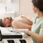
- A transoesophageal echocardiogram (TOE/TEE) passes a small ultrasound probe into the oesophagus, the food pipe, to image the heart from directly behind it, providing far higher resolution than a standard chest-wall echo.
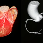
- The oesophagus sits immediately behind the heart, allowing the probe to capture structures that are difficult or impossible to see clearly from the chest wall, particularly the mitral valve, left atrium, and aorta.
- The procedure is performed under sedation with throat numbing spray. You will be comfortable and relaxed throughout and will have no memory of the probe being passed.
- Fasting for at least 6 hours beforehand is essential. You will need someone to take you home, you cannot drive after sedation.
- A mild sore throat for a day or two afterwards is common and expected. Serious complications are rare.
For most patients, a standard echocardiogram through the chest wall provides everything a cardiologist needs. But the heart has a posterior surface, structures at the back, that the chest-wall probe cannot always see clearly. The mitral valve, the left atrium, the left atrial appendage, and portions of the aorta all sit in this zone.
When those structures are the clinical question, a transoesophageal echocardiogram, TOE in Australian and British practice, TEE in North American, is the investigation that answers it.
The idea of a probe being passed into the oesophagus understandably makes patients anxious. The reality is considerably more comfortable than the description suggests, and understanding exactly what happens helps take the anxiety out of the procedure well before you arrive for it.
Why the Oesophagus?
The oesophagus, the food pipe running from the throat to the stomach, passes directly behind the heart. Placing an ultrasound probe inside it puts the transducer within centimetres of the cardiac structures, with no chest wall, ribs, or lung tissue in the way.
The result is image resolution and detail that simply cannot be achieved from the chest wall. Structures that appear blurred or partially obscured on a standard echo are seen with clarity on a TOE. For clinical decisions that depend on precise anatomical information, valve repair planning, clot detection, infective endocarditis assessment, this detail changes everything.
When Is a TOE Requested?
The most common indications
Valve disease assessment
Detailed assessment of the mitral valve, particularly before repair or replacement surgery, where precise anatomy determines the surgical approach.
Blood clot detection
Detection of clots in the left atrial appendage, critical before cardioversion of AF, and in stroke investigation.
Infective endocarditis
Detection of vegetations, infected growths on heart valves, which can be missed on a standard chest-wall echo.
Structural defects
Assessment of septal defects, ASD, PFO, including precise sizing for catheter-based closure procedures.
Aortic disease
Assessment of the thoracic aorta for dissection, aneurysm, or atherosclerotic plaque, structures that lie adjacent to the oesophagus.
Procedural guidance
Real-time imaging during cardiac procedures, valve repair, TAVI, septal closure, to guide catheter positioning and confirm results.
What to Expect
What to Expect, Transoesophageal Echocardiogram (TOE/TEE)
Duration
The imaging itself takes 20–40 minutes. With preparation and recovery, allow 2–3 hours for the appointment.
Preparation
Fast for at least 6 hours beforehand, nothing to eat or drink except water. Arrange for someone to take you home. You cannot drive after sedation.
Comfort
A sedative is given intravenously and the throat is numbed with a local anaesthetic spray before the probe is passed. Most patients have little or no memory of the procedure and wake feeling comfortable.
Radiation / Contrast
No radiation. Ultrasound only. No iodine contrast dye is required.
Results
The cardiologist reviews images immediately. A formal report is typically available within a few days and discussed at follow-up or, where urgent, before you leave.
Afterwards
Wait until throat numbness has fully worn off before eating or drinking, usually 1–2 hours. A mild sore throat for a day or two is common. Rest for the remainder of the day.
The Procedure, Step by Step
Before the probe is passed
When you arrive, a cannula is placed in a forearm vein for the sedation. ECG leads and a blood pressure cuff are applied, and oxygen saturation is monitored throughout. A throat numbing spray is applied to the back of the throat, this reduces the gag reflex and any discomfort during probe passage. The sedative is then given through the cannula.
The sedation used for a TOE is not general anaesthesia, it is a short-acting agent that produces deep relaxation, drowsiness, and often amnesia for the procedure itself. You remain breathing independently throughout. Most patients describe waking up and being surprised it is already over.
The imaging
The probe is a slim, flexible tube approximately the diameter of a finger, with a small ultrasound transducer at the tip. Once sedation takes effect and the gag reflex is suppressed, it is gently guided over the back of the tongue and into the oesophagus. This is done carefully and takes only a moment.
The cardiologist then manoeuvres the probe to obtain images from multiple positions within the oesophagus and stomach, each position revealing different structures and angles of the heart. The imaging itself is typically complete within 20 to 30 minutes.
Recovery
After the probe is removed you are monitored in a recovery area while the sedation wears off. This typically takes 30 to 60 minutes. Your throat will remain numb for a period, do not eat or drink until the numbness has fully resolved, to avoid the risk of choking. A mild sore throat over the following day or two is expected and resolves on its own.
The most common thing patients say to me after a TOE is that it was far less unpleasant than they expected. The sedation is effective, the throat spray works well, and the whole experience passes quickly. I encourage patients to be honest about their anxiety beforehand, we can always adjust the sedation to ensure they are comfortable.
Risks and Safety
A TOE is a safe procedure in the vast majority of patients. The most common side effect is a temporary sore throat. Serious complications, oesophageal injury, significant bleeding, or a severe reaction to sedation, are uncommon and the procedure is performed in an environment equipped to manage them.
Patients with known oesophageal conditions, strictures, severe dysmotility, or previous oesophageal surgery, should inform the team, as these may affect suitability for the procedure. Your cardiologist will have considered this before making the referral.
- Why is a TOE needed rather than a standard echocardiogram, what specifically is being looked for?
- I have an oesophageal condition, does this affect the procedure?
- How much sedation will I be given, and will I be aware during the procedure?
- Can I take my regular medications on the day, or do any need to be withheld?
- How quickly will the results be available and who will discuss them with me?
Heart Matters Resource
When in Doubt, Get Checked Out
If your cardiologist has recommended a TOE, it is because they need a level of detail that a standard echocardiogram cannot provide. The anxiety about the procedure is understandable, but the information it gives is worth having.
Conclusion
The transoesophageal echocardiogram is the investigation that answers the questions a standard echo cannot. When precise detail of the mitral valve, the left atrium, the left atrial appendage, or the aorta is clinically necessary, for diagnosis, for treatment planning, or for procedural guidance, a TOE provides that detail reliably and safely.
The procedure is considerably more comfortable than patients typically expect. The sedation is effective, the throat spray is well-tolerated, and most patients have little memory of it by the time they are in recovery. Understanding what is involved, and why it has been requested, is the best preparation.
If you have questions or concerns before your procedure, raise them with your team in advance. The more information you have, the less anxiety you will carry into the appointment.