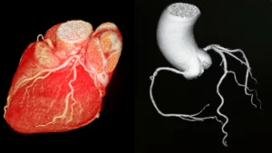
- An echocardiogram, or “echo”, is an ultrasound of the heart. It uses sound waves to create moving, real-time images of the chambers, valves, walls, and blood flow.
- It is the single most important imaging test in cardiology for assessing how the heart is built and how well it pumps, measuring ejection fraction, valve function, wall thickness, and chamber size.
- It is completely non-invasive and painless, uses no radiation, and takes around 30 to 45 minutes.
- It is requested across a wide range of situations, from investigating a murmur or breathlessness to monitoring heart failure, checking the heart before surgery, or assessing recovery after a heart attack.
- Specialised forms include the stress echocardiogram and the transoesophageal echocardiogram (TOE/TEE), both covered in their own articles in this section.
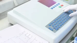
If the ECG is the heart’s electrical portrait, the echocardiogram is its structural one. Where an ECG tells your cardiologist about rhythm and electrical signals, the echo shows the physical heart itself, how large the chambers are, how the walls move with each contraction, whether the valves open and close properly, and how blood flows through the heart.
It answers questions about structure and function that no other test addresses as completely, as safely, or as gently. For many patients, it is the investigation that finally explains their symptoms.
How It Works
Sound waves, not radiation
An echocardiogram uses the same ultrasound physics as a pregnancy scan. High-frequency sound waves are sent from a small handheld probe, bounce back from the structures inside the chest, and are turned into moving images on a monitor. No radiation is involved at any point.
A cardiac sonographer places the probe on the skin of the chest, using a clear gel to remove the air gap between the probe and the skin, because air blocks the sound waves. By moving the probe to different positions around the chest, several viewing angles are captured, building a thorough picture of every part of the heart.
Doppler imaging, measuring blood flow
Modern echocardiography also uses Doppler imaging, which measures the speed and direction of blood as it flows through the heart. This lets your cardiologist judge how severe any valve narrowing or leaking is, track changes over time, and decide whether and when treatment such as surgery might be needed.
What an Echo Can Show
An echocardiogram provides several different kinds of information at the same time, which is part of what makes it one of the most useful single tests in cardiology.

Key measurements and findings
Ejection fraction
The percentage of blood pumped out with each beat, normally above 55%. It is the main measure of how well the heart squeezes.
Chamber dimensions
Whether the heart is enlarged in response to extra volume or pressure, which helps guide diagnosis and treatment decisions.
Valve function
All four valves are checked for stenosis (narrowing) and regurgitation (leaking), with Doppler measuring how severe each one is.
Wall motion
Areas of the heart muscle that contract poorly can point to a previous heart attack, reduced blood supply, or cardiomyopathy.
Pericardial effusion
Fluid around the heart can be spotted straight away and measured. It can point to pericarditis or other conditions.
Structural findings
Congenital defects present from birth, cardiac masses, and other structural issues are often picked up first on a routine echo.
What to Expect
What to Expect, Transthoracic Echocardiogram (TTE)
Duration
30 to 45 minutes for a full study. A brief, focused scan in an emergency may take 10 to 15 minutes.
Preparation
No fasting or special preparation is needed. Wear comfortable clothing that allows easy access to the chest.
Comfort
Painless and non-invasive. You lie on your left side for much of the scan, which moves the heart closer to the chest wall for clearer images. The gel feels cold at first. Sometimes firmer pressure with the probe is needed to get a good view.
Radiation / Contrast
No radiation, ultrasound only. For some scans, a tiny-bubble ultrasound contrast may be injected to sharpen the images. This is not iodine contrast and is different from CT contrast.
Results
A cardiologist interprets the recorded images. A formal report is usually ready within a few days and discussed at your follow-up.
Afterwards
No restrictions. You can drive home and return to normal activities straight away. The gel washes off easily.
Why Your Cardiologist Has Requested One
The echocardiogram is one of the most widely requested tests in cardiology, because information about how the heart is built and how well it works matters across so many different situations.
Common reasons include investigating a heart murmur heard through the stethoscope, assessing breathlessness where a cardiac cause is possible, setting a baseline and monitoring known valve disease or heart failure, checking heart function after a heart attack, assessing the heart before surgery, looking into atrial fibrillation to measure left atrial size and ventricular function, investigating a suspected cardiomyopathy, and monitoring the heart during certain cancer chemotherapy treatments.
The echocardiogram often gives me the single most useful piece of information in a heart assessment. Knowing the ejection fraction, the valve function, and the chamber sizes at once, in real time, with no radiation and nothing invasive, is remarkable. It is the test I rely on most.
Associate Professor Nagesh Anavekar, Cardiologist
Types of Echocardiogram
Standard transthoracic echocardiogram (TTE)
The most common form, with the probe placed on the chest wall. This is the starting point for most echocardiography and the scan described in this article.
Stress echocardiogram
This combines a standard echo with exercise on a treadmill or bicycle, or with a medication that mimics exercise, to see how the heart copes with extra demand. It is used mainly to look for coronary artery disease and to assess valves under load. We have a dedicated article on the stress echocardiogram in this section.
Transoesophageal echocardiogram (TOE/TEE)
This passes the probe gently into the oesophagus to get clearer images of structures at the back of the heart, particularly the mitral valve, the left atrium, and the aorta. It is used when a standard echo cannot show enough detail, or when very high-resolution images are needed to plan a procedure. It is covered in our dedicated article on the transoesophageal echocardiogram.
- What is my ejection fraction, and what does that number mean for me?
- Were any valve problems found, and if so, how significant are they?
- Will I need a repeat echo, and if so, how often?
- Is a stress echo or a TOE needed to give more information?
- What did the echo show that my ECG or other tests could not?
Heart Matters Resource
When in Doubt, Get Checked Out
Breathlessness, a heart murmur, palpitations, or chest discomfort that has not been explained. An echocardiogram is often one of the first and most informative steps towards an answer.
Conclusion
The echocardiogram is one of cardiology’s most versatile and informative tests. It answers questions about structure, function, and blood flow that no blood test or ECG can provide, with no radiation and nothing invasive required.
Whether it is confirming a normal ejection fraction in someone worried about their heart, measuring the severity of valve disease that needs watching, or finding wall motion changes after a heart attack, the information it provides shapes real decisions about your care. If you have had an echo and are unsure what the report means, the questions box above is a good place to start your next conversation with your cardiologist.
Related Reading
- The Stress Echocardiogram: What It Is, What to Expect, and What the Results Mean
- Transoesophageal Echocardiogram (TOE/TEE): What It Is and What to Expect
- The Electrocardiogram (ECG / EKG)
- Troponin: The Blood Test at the Heart of Chest Pain Assessment
- CT Coronary Angiogram (CTCA): What It Is, What to Expect, and What It Shows
- Heart Failure With Reduced Ejection Fraction: Why Quadruple Therapy Changed Everything
- Understanding Your Cardiovascular Risk Factors