- Pericarditis is inflammation of the thin sac surrounding the heart. It is one of the more common causes of chest pain in younger people and, in most cases, one of the least dangerous.
- The pain has a distinctive signature. It typically sharpens when lying flat and eases on sitting forward, which is unusual enough to be a useful clue.
- In most cases no cause is ever found, or the illness follows an ordinary virus. Where tuberculosis is common, it is a leading cause and the picture differs. Where a specific cause does turn up, it changes both the outlook and the treatment.
- Anti-inflammatory treatment combined with colchicine is standard first-line practice. In trial data, adding colchicine roughly halved the chance of the condition returning.
- Recurrence is the most common complication, affecting somewhere between 15% and 30% of people after a first episode. Serious complications are uncommon. Where no underlying cause is found, fewer than 1 in 100 people go on to develop lasting stiffening of the sac.
The pain arrives sharply, usually behind the breastbone, and it behaves in a way that heart pain is not supposed to behave. Lying down makes it worse. Sitting up and leaning forward makes it better. Taking a deep breath can be enough to catch it.
That pattern sends many people to an emergency department convinced they are having a heart attack, and it is also why the diagnosis is often made quickly once someone describes it. Few cardiac symptoms are as positionally specific.
It tends to affect people younger than the typical coronary patient, and the outlook for most is good. What follows is what the condition actually is, why it happens, and what the evidence supports doing about it.
What the Pericardium Does
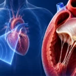
The heart sits inside a two-layered sac called the pericardium. Between the layers is a small amount of fluid, usually only 15 to 50 mL, which allows the heart to move smoothly against surrounding structures with every beat.
The sac anchors the heart, provides a barrier against infection spreading from the lungs, and stops the heart stretching too far. It is not essential to survival, and people who have had it surgically removed generally do well.
When it becomes inflamed, two things follow. The layers lose their smooth glide and rub against each other, which produces pain. Inflammation can also draw extra fluid into the space between the layers. Doctors call that a pericardial effusion, which simply means a build-up of fluid around the heart.
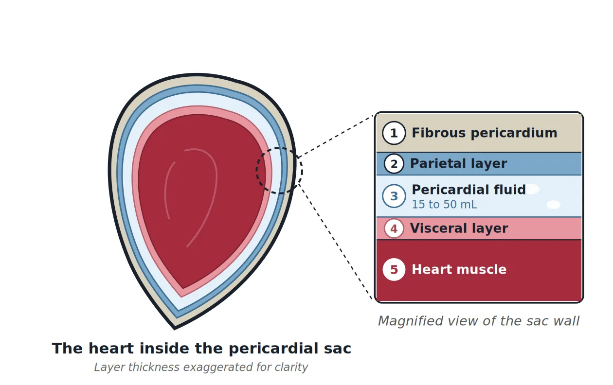
Fibrous pericardium, the outer layer. The tough outside of the sac. It anchors the heart in place and stops it stretching too far.
Parietal layer, the outer lining. The smooth inside surface of the outer sac.
Pericardial fluid. Normally only 15 to 50 mL, enough for the surfaces to glide. Inflammation can increase it, and the build-up is called an effusion.
Visceral layer, the inner lining. The same thin membrane as layer 2, folded back on itself and stuck to the heart’s own surface. Its medical name is the epicardium.
Heart muscle. Sits beneath the sac and is not affected in straightforward pericarditis. Doctors call it the myocardium.
What It Feels Like
The pain has a recognisable pattern
The pain is sharp rather than heavy, sits centrally behind the breastbone, and often radiates to the neck, the left shoulder, or the ridge of muscle running along the top of the shoulder. That last spot is unusual, and when it happens it is a fairly strong clue.
What sets it apart is how it responds to position and breathing. Lying flat brings the inflamed layers into closer contact and intensifies the pain. Sitting upright and leaning forward relieves it. Deep breaths, coughing, or swallowing often make it worse.
This is close to the opposite of the pain of a heart attack, which is typically a pressure or heaviness, unaffected by position, and frequently accompanied by sweating and nausea. The distinction is useful but not absolute, which is why chest pain of any kind is assessed rather than self-diagnosed.
Other symptoms
A low-grade fever is common, particularly where a viral illness preceded the episode. Fatigue, breathlessness, and a general sense of being unwell frequently accompany the pain, and symptoms tend to build over hours rather than striking instantaneously.
How the Diagnosis Is Made
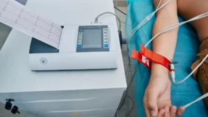
European guidelines define acute pericarditis by the presence of at least two of four features: the characteristic chest pain, a particular sound heard through the stethoscope, specific changes on the heart tracing, and new or increasing fluid around the heart.
That sound has a name, a pericardial friction rub, and it is exactly what it sounds like: a scratching or creaking made by the inflamed layers rubbing together. Hearing it points strongly to the diagnosis. It comes and goes, though, so not hearing it rules nothing out.
No single test confirms it. The diagnosis is assembled from several, each answering a different question.
| Test | What it looks for | In pericarditis |
|---|---|---|
| ECG | The heart’s electrical signal | Widespread, not one artery |
| Echocardiogram | Fluid around the heart | Effusion, filling affected |
| CRP | Inflammation in the body | Raised, tracks response |
| Troponin | Damage to heart muscle | Raised if muscle involved |
| Cardiac MRI | Inflammation, seen directly | Used when unclear |
The ECG point is the one that separates pericarditis from a heart attack most reliably. Its changes are widespread rather than confined to the territory of a single artery. Troponin matters for a different reason. It is a protein released when heart muscle is damaged, so a raised level means the muscle is inflamed alongside the sac. That combination has its own name, myopericarditis, and it is watched more closely.
What Causes It
Most often, no cause is ever found
Where tuberculosis is uncommon, most cases fall into one of two groups: no cause found, or an ordinary virus assumed to be behind it. Doctors label the first group idiopathic, which is simply the medical word for cause unknown.
The two groups blur into each other. Many of the unexplained cases were almost certainly viral all along, with no test ever done to pin down which virus, largely because finding out rarely changes the treatment.
The viruses involved are everyday ones: the family behind common colds and stomach upsets, influenza, and the virus that causes glandular fever. A chest or stomach bug in the few weeks beforehand is a common part of the story.
That is why guidelines in low-tuberculosis regions do not push for exhaustive testing in every case: the search seldom finds anything and the usual causes settle on their own. Where tuberculosis rates are high, it becomes the leading cause of pericarditis and the approach changes completely.
COVID-19 and the vaccine question
This deserves a direct answer rather than a careful silence, because it is what many readers arrive looking for.
Both COVID-19 itself and the mRNA COVID vaccines have been linked to inflammation of the sac and of the heart muscle. Both associations are real. They differ substantially in magnitude.
Analyses of national reporting systems have estimated roughly six cases of heart or sac inflammation per million vaccine doses given, across all ages. Risk concentrates heavily in one group: young males, after the second dose, with estimates of around 12 to 13 cases per million doses in those aged 12 to 39. In adolescent studies, the risk after a second dose has been several times higher than after a first, and considerably higher in males than females. Most cases have been mild and settled with standard treatment.
Catching the virus carries the higher risk. A Hong Kong population study found this inflammation in roughly 326 per million people with confirmed COVID-19, against about 5.5 per million among those vaccinated, the latter being no different from the pre-pandemic background rate. A Spanish hospital study reported the same direction of effect, with the incidence following infection around 20 times that following mRNA vaccination. Cases attributed to infection also tended to be more severe.
Estimates vary between studies according to population, age structure, case definition, and how thoroughly cases were sought, so the precise figures should be read as an order of magnitude rather than a fixed number. The consistent finding across countries is the direction of the comparison. The risk is real on both sides, is counted in cases per million on both sides, and is higher after infection than after vaccination.
None of this makes an individual case less real or less unpleasant for the person experiencing it. Someone who develops pericarditis after a vaccine dose has a genuine condition requiring genuine treatment, and population-level reassurance is not an answer to an individual’s symptoms.
The causes at a glance
No cause found
Nothing is ever pinned down. This is the biggest single group, and it is also the one that settles most easily. Doctors call it idiopathic.
Viral illness
Everyday viruses: colds, stomach bugs, influenza, glandular fever. A chest or stomach illness in the previous few weeks is a common part of the story.
COVID and vaccination
Both infection and mRNA vaccination have been linked to it. Both are counted in cases per million, and the figure is higher after infection. See the section above.
Injury to the heart
After a heart attack, heart surgery, or a procedure done through a tube in an artery. The sac reacts to the injury nearby.
Autoimmune and systemic
Conditions where the immune system attacks the body’s own tissues, such as lupus and rheumatoid arthritis. Also kidney failure, some cancers, radiation treatment to the chest, and certain medicines.
Bacterial and tuberculous
Bacterial infection or tuberculosis. Far less frequent where tuberculosis rates are low, but wherever it occurs the outlook is considerably worse and lasting stiffening of the sac is far more likely.
How It Is Treated
Clinicians work through a fairly settled sequence. Most people never get past the first step of it.
| Treatment | Where it fits | Typical duration |
|---|---|---|
| Aspirin or ibuprofen | First choice, for the pain | Until symptoms settle |
| Colchicine | Added to the first choice | 3 months, 6 if it returns |
| Steroid tablets | Where the first two are unsuitable | Reduced slowly |
| Anakinra, rilonacept | Repeated relapses only | Specialist supervision |
Dose and duration are individual. Only the prescribing doctor, who knows the specific circumstances, can determine what is appropriate for a given person.
Two things in that table are worth expanding on.
Colchicine earns its place. The evidence is unusually strong for a condition this common. In the ICAP trial, people having a first episode were randomly given either colchicine or a dummy tablet alongside their usual treatment. The illness persisted or came back in 16.7% of those on colchicine, against 37.5% on the dummy tablet. Looking at return of symptoms alone, the figures were 9.2% against 20.8%. Stomach and bowel side effects were no more common than with the dummy tablet.
Steroids sit third for a reason. They work quickly, which makes their position in the list counter-intuitive. But steroids used during a first attack have been linked to a higher chance of the problem returning, so the medicine that settles things fastest can make the months afterwards harder. They are generally kept for people who cannot take the first two options, or where an immune condition is being treated, and only once infection has been ruled out.
Under 1% get a stiff sac
Where the cause is unknown or viral. The figure rises sharply where bacteria or tuberculosis are responsible.
15% to 30% recur
Recurrence is the most common complication after a first episode, and the figure is higher again without colchicine.
Colchicine halves recurrence
In the ICAP trial, the illness came back or persisted in 16.7% on colchicine, against 37.5% on a dummy tablet.
When It Comes Back
Recurrence is the most common complication and the most frustrating part of the condition. Between 15% and 30% of people experience a recurrence after a first episode, and among those who go on to have a first recurrence, the chance of a further one rises towards 50%.
A recurrence means a fresh episode after four to six weeks with no symptoms at all. The best explanation is that the immune system keeps the reaction running long after whatever set it off has gone. That is why medicines aimed at the immune signal itself work when they do.
The reassuring part, and it is worth stating plainly because people with repeated episodes often assume the opposite, is that recurrence does not signal accumulating damage. In groups of patients followed through repeated unexplained episodes, lasting stiffening of the sac has not been reported at all, and the overall risk in this group looks lower than after a single first episode.
The Complications That Matter
Two complications account for most of the concern.
Fluid squeezing the heart. If fluid gathers quickly enough, or in a large enough volume, it presses on the heart and stops it filling properly. The medical name is cardiac tamponade. It is rare where no cause is found, and much more common where cancer, tuberculosis or bacterial infection is behind the illness. Treatment is to drain the fluid.
A sac that turns stiff. Long-running inflammation can leave the sac thickened and rigid, so it grips the heart like a shell and limits every beat. The medical name is constrictive pericarditis. The risk is under 1% where the cause was unknown or viral, 2% to 5% for immune and cancer-related causes, and 20% to 30% where bacteria or tuberculosis were responsible.
Signs that warrant urgent medical attention include fainting, rapid or laboured breathing, severe or escalating breathlessness, or a sudden deterioration. These can be signs of fluid building up and squeezing the heart. Anyone experiencing them should seek emergency care immediately rather than waiting for a scheduled appointment.
Conclusion
Pericarditis is a condition where the alarming presentation and the actual outlook are poorly matched. The pain is genuinely severe, it mimics a heart attack closely enough to send people to hospital, and yet the great majority of cases settle with anti-inflammatory treatment and leave nothing behind.
Two things most influence how it goes: whether an underlying cause is identified, which changes the outlook substantially, and whether colchicine forms part of the initial treatment, which roughly halves the chance of it returning. For a diagnosis that so often arrives frightening, that is an encouraging picture.
Related Reading
- Chest Pain: Understanding Symptoms, Causes and Evaluation
- Inflammation and Heart Disease: The Hidden Driver Behind Atherosclerosis, Pericarditis, and More
- The Electrocardiogram (ECG / EKG)
- The Echocardiogram: What It Shows and Why It Matters
- What Is a Troponin Test, and Why Is It Used for Chest Pain?
- Heartburn or Heart Attack? How to Tell the Difference
- Chest Tightness and the Heart: Why This Symptom Demands Attention