The Pulse Newsletter Expert heart health, in plain English. Fortnightly, from our clinicians. Subscribe →

The Nuclear Stress Test: What It Is, What to Expect, and Why It’s Requested

Key Points

  • A nuclear stress test, also called myocardial perfusion imaging (MPI), uses a small amount of radioactive tracer to produce detailed images of blood flow through the heart muscle at rest and during stress.
  • It shows not just whether the heart is receiving adequate blood supply, but precisely which areas of muscle are affected and to what degree, making it particularly useful for planning treatment after a heart attack or assessing complex coronary disease.
  • The radiation dose is low and the tracer clears from the body within hours. The test is safe and well-tolerated.
  • For patients who cannot exercise, a pharmacological stress agent is used to mimic the effect of exertion on the heart, making the test accessible regardless of physical capacity.
  • The nuclear stress test is typically requested when other investigations have not provided sufficient information, or when a more detailed map of myocardial perfusion is needed for clinical decision-making.

Most people who are investigated for chest pain or breathlessness will have a stress echocardiogram or CT coronary angiogram as their functional or anatomical assessment. But there is a subset of clinical questions where a more detailed map of blood flow through the heart muscle, not just whether flow is reduced, but precisely where and by how much, changes the clinical decision. That is where myocardial perfusion imaging comes in.

The nuclear stress test is less commonly performed than other cardiac investigations, but when it is requested it is usually for a specific and well-considered reason. Understanding what it involves and what it provides helps patients approach it with appropriate expectations.

What Is a Nuclear Stress Test?

Myocardial perfusion imaging

A nuclear stress test, formally called myocardial perfusion imaging (MPI) or radionuclide stress testing, uses a small amount of a radioactive tracer injected into a vein. This tracer travels through the bloodstream and is taken up by the heart muscle in proportion to blood flow. Areas receiving good blood supply absorb more tracer; areas with reduced flow absorb less.

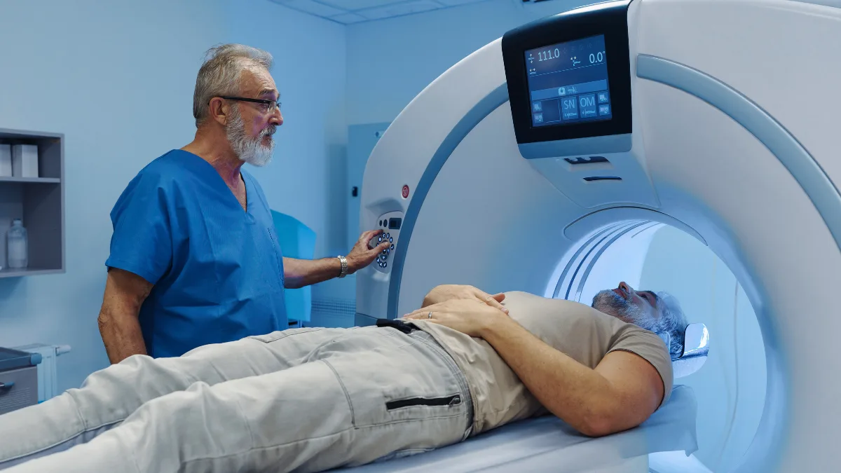
A specialised camera, called a gamma camera or SPECT scanner, then detects the radiation emitted by the tracer and produces detailed images of the distribution of blood flow through the heart muscle. Images are taken at rest and during stress, and the two sets are compared.

What it can show

Where a stress echocardiogram detects wall motion abnormalities as an indirect consequence of reduced blood flow, myocardial perfusion imaging detects the reduced flow directly. This makes it particularly sensitive for identifying ischaemia, reduced blood supply to the heart muscle during stress, and for mapping its extent and distribution with precision.

It can also distinguish between viable heart muscle that is simply underperfused, and therefore potentially recoverable, and permanently scarred tissue from a previous heart attack that will not recover regardless of intervention. This distinction is clinically important when deciding whether revascularisation is likely to benefit a patient.

When Is It Requested?

The nuclear stress test occupies a specific niche in the cardiac investigation toolkit. It is typically requested in clinical situations where other investigations have not provided sufficient information, or where the level of anatomical and functional detail it provides is specifically needed.

Common indications include assessment of known coronary artery disease where the functional significance of a narrowing needs to be established; evaluation of myocardial viability after heart attack to determine whether revascularisation is likely to improve function; investigation of chest symptoms in patients with complex anatomy, such as previous bypass surgery, where standard stress testing may be less reliable; and risk stratification in patients with known coronary disease prior to non-cardiac surgery.

What to Expect, Nuclear Stress Test

Duration

3–4 hours in total across the appointment, including rest imaging, stress phase, and post-stress imaging. Some protocols split rest and stress imaging across two days.

Preparation

Fast for 4–6 hours before the test. Avoid caffeine for 24–48 hours, caffeine interferes with pharmacological stress agents. Some medications may need to be withheld, your team will advise specifically. Wear comfortable clothing and walking shoes.

Comfort

A cannula is placed in a vein in the arm for the tracer injection. The imaging involves lying still on a scanner table with the camera rotating around the chest, this is not enclosed and is well-tolerated. The pharmacological stress agent may cause a brief flushing sensation or mild breathlessness that resolves quickly.

Radiation

A small amount of radioactive tracer is used. The effective radiation dose is typically 3–10 mSv, comparable to a CT coronary angiogram. The tracer clears from the body within hours to a day. No special precautions are required afterwards.

Results

Images require specialist nuclear cardiology reporting. Results are typically available within a few days and discussed with you by your referring cardiologist at follow-up.

Afterwards

No restrictions on activity. Drink plenty of water to help the tracer clear. If you had a pharmacological stress agent, any transient symptoms resolve quickly, you will be monitored until they do.

Exercise vs Pharmacological Stress

Exercise stress

Where possible, physical exercise on a treadmill or exercise bike is the preferred method of inducing stress, it produces the most physiologically meaningful assessment and also provides additional information about exercise capacity, heart rate response, and blood pressure behaviour during exertion.

Pharmacological stress

For patients who cannot exercise adequately, due to orthopaedic limitations, severe deconditioning, peripheral vascular disease, or other reasons, a pharmacological stress agent is used instead. Adenosine, regadenoson, or dobutamine are the most commonly used agents. They work by dilating the coronary arteries or increasing heart rate and myocardial demand, mimicking the effect of exercise on blood flow distribution.

Caffeine blocks the effect of adenosine-based agents, which is why avoiding coffee, tea, and other caffeine sources for 24 to 48 hours before the test is essential if a pharmacological stress protocol is planned. Your team will confirm the specific requirements.

How It Compares to Other Tests

Test What it shows Radiation Best used for
Stress echocardiogram Wall motion, indirect marker of ischaemia None First-line functional assessment, valve assessment under load
CT coronary angiogram Coronary artery anatomy, degree of narrowing Low (3–10 mSv) Ruling out significant coronary disease, anatomical planning
Nuclear stress test (MPI) Blood flow distribution through heart muscle Low–moderate (3–10 mSv) Detailed perfusion mapping, viability assessment, complex CAD
Cardiac MRI stress test Perfusion and function, no radiation None Detailed perfusion without radiation, increasingly used
Invasive coronary angiogram Direct coronary anatomy, gold standard Low (radiation from X-ray) Definitive diagnosis with option to treat in same session

About the Radiation

The word “nuclear” in nuclear stress test refers to the radioactive tracer, not to the type of energy or any risk analogous to nuclear power. The tracers used are specifically designed for rapid clearance from the body and produce a radiation dose that, while slightly higher than a CT coronary angiogram, remains well within accepted safety parameters for a single diagnostic test.

To put it in context, the effective dose from a nuclear stress test is roughly equivalent to one to three years of natural background radiation from the environment. The clinical benefit of accurate diagnosis in the situations where a nuclear stress test is requested far outweighs this level of exposure.

When I request a nuclear stress test, it is usually because I want more information than other investigations have provided, specifically, I want to know not just whether there is ischaemia, but where it is and how much muscle is affected. That precision changes decisions. It can be the difference between recommending revascularisation and recommending optimised medical therapy.

— Prof. Peter Barlis, Interventional Cardiologist

Questions worth asking about your nuclear stress test

  • Why is a nuclear stress test being requested rather than a stress echo or CTCA, what specifically does it add?
  • Will I be exercising or having a pharmacological stress agent, and does caffeine avoidance apply to me?
  • Which medications do I need to withhold before the test?
  • What will the results tell you that you don’t already know from my other investigations?
  • Is cardiac MRI an alternative that could provide similar information without radiation?

Heart Matters Resource

When in Doubt, Get Checked Out

If you have been referred for a nuclear stress test and want to understand why, or what the result will mean for your management, that conversation with your cardiologist before the test is worth having.

Read: When in Doubt, Get Checked Out →

Conclusion

The nuclear stress test is a specialised investigation that provides a level of detail about myocardial blood flow that other non-invasive tests cannot match. When the clinical question requires knowing not just whether ischaemia is present, but where and how much muscle is affected, or whether damaged heart muscle is still viable, myocardial perfusion imaging provides the answer.

The radiation involved is modest and the procedure is well-tolerated. The length of the appointment, typically three to four hours, is the main practical consideration, and knowing to expect this in advance makes the day considerably less uncertain.

If you have been referred for this test, it is because your cardiologist needs a specific type of information to guide your management. That information will shape the next clinical decision, and that is exactly what a good investigation should do.
More from Heart Matters, Diagnostic Tests

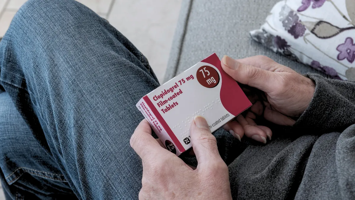
What Is Clopidogrel Used For?

heartmatters.com 2026 04 02T052736.709

Clopidogrel is one of the most widely prescribed heart medicines in the world. Here is how it works, what it is used for, and everything you need to know about taking it safely.

Is There Something Fishy About Fish Oil?

heartmatters.com 2026 04 05T090155.280

A recent study links regular fish oil supplements to increased cardiovascular risk in people without pre-existing heart disease. Here is what the findings mean for you.

Shortness of Breath and the Heart: When Breathlessness Is a Cardiac Signal

heartmatters.com 2026 03 31T205341.285
Key Points

  • Shortness of breath, breathlessness, is one of the most common reasons people see a cardiologist, and one of the most diagnostically important symptoms in cardiovascular medicine.
  • The heart and lungs work as a unit. When the heart is not pumping efficiently, fluid backs up into the lungs, producing breathlessness that is often the first signal something is wrong.
  • Breathlessness on exertion that is new, progressive, or out of proportion to effort deserves cardiac assessment, even if it seems to have an obvious non-cardiac explanation.
  • A normal BNP blood test and a normal echocardiogram together substantially reduce the likelihood of a cardiac cause and redirect the investigation efficiently.
  • Breathlessness at rest, particularly when lying flat, waking you from sleep, or accompanied by chest discomfort, requires prompt assessment, not a wait-and-see approach.

Breathlessness is such a common human experience, after exercise, after a shock, at altitude, that it is easy to normalise it when it shouldn’t be. Patients often come to me having been breathless for months, having attributed it to being unfit, getting older, putting on weight, or stress.

Sometimes those explanations are correct. But breathlessness is also one of the most important cardiac symptoms there is, and one of the most frequently under-investigated. The heart and lungs are inseparable in their function, and when the heart begins to struggle, the lungs are often the first place it shows.

Understanding what cardiac breathlessness feels like, what causes it, and when to seek assessment is genuinely important, because the earlier a cardiac cause is identified, the better the options for treatment.

Why the Heart Causes Breathlessness

The heart-lung connection

The left side of the heart receives oxygenated blood from the lungs and pumps it out to the body. When the left ventricle is not pumping efficiently, whether from heart failure, a weak muscle, a valve problem, or elevated pressures, blood backs up through the pulmonary circulation. Fluid accumulates in the lung tissue itself, making the lungs stiffer and gas exchange less efficient.

The result is breathlessness, the sensation of not being able to get enough air in, of breathing being harder than it should be. At rest this may be subtle or absent. With exertion, when the heart needs to increase its output and cannot do so adequately, the breathlessness becomes apparent.

Why it so often gets missed

The insidious onset of cardiac breathlessness is part of why it gets missed. It rarely arrives suddenly, it creeps in gradually, week by week. A person who used to walk briskly up a hill now walks slowly. Someone who carried groceries up the stairs now takes two trips. The brain unconsciously recalibrates what feels normal, and by the time the breathlessness is significant enough to seek medical attention, a meaningful period of cardiac stress may have already accumulated.

One of the most telling questions I ask in clinic is: “What could you do six months ago that you can’t do now?” That question unlocks the real story, because patients adapt so well to progressive breathlessness that they often don’t realise how much their functional capacity has changed until someone asks directly.

Cardiac Causes of Breathlessness

Heart failure

Heart failure, reduced pump function of the left ventricle, often expressed as a reduced ejection fraction, is the most common cardiac cause of breathlessness. The failing heart cannot maintain adequate output, pressures rise in the pulmonary circulation, and fluid accumulates in the lungs. Breathlessness on exertion is the hallmark symptom, often accompanied by fatigue, ankle swelling, and reduced exercise tolerance.

Heart failure with preserved ejection fraction (HFpEF), where the pumping function appears normal but the heart muscle is stiff and fills abnormally, is increasingly recognised and produces breathlessness through a subtly different mechanism, but the symptom pattern is similar.

Valve disease

Significant valve disease, particularly aortic stenosis and mitral regurgitation, can produce breathlessness as the heart compensates for abnormal flow across the valve. Aortic stenosis in particular can progress for years without symptoms, then produce breathlessness, chest pain, or fainting as the valve area becomes critically narrow. An echocardiogram identifies valve problems with precision.

Atrial fibrillation

AF causes breathlessness in two ways. The irregular rhythm and often elevated heart rate reduce the efficiency of cardiac filling and output, particularly in people whose hearts rely on the atrial contraction component that AF abolishes. Additionally, AF is often a manifestation of underlying cardiac disease that itself causes breathlessness. Many people first notice their AF through unexplained breathlessness rather than palpitations.

Coronary artery disease

Significant narrowings in the coronary arteries can cause breathlessness as an anginal equivalent, particularly in women, in people with diabetes, and in older adults, where classic chest pain may be absent or minimal. Breathlessness on exertion that resolves with rest, in someone with cardiovascular risk factors, should always raise the possibility of ischaemia.

Pulmonary hypertension

Elevated pressure in the pulmonary circulation, whether from left heart disease, lung disease, or primary pulmonary arterial hypertension, causes breathlessness that can be severe and progressive. It is a diagnosis that requires specialist assessment but is important not to miss, particularly in younger patients with breathlessness that seems disproportionate to their apparent health.

Non-Cardiac Causes, Important to Distinguish

Not all breathlessness is cardiac, and part of the clinical task is efficiently distinguishing between causes. The most common non-cardiac causes include asthma and COPD, pulmonary embolism (blood clot in the lungs), anaemia, obesity, deconditioning, anxiety and panic disorders, and thyroid disease.

Several of these can coexist with cardiac disease, which is why the investigation is rarely a matter of ruling out one thing, it is a matter of understanding which factor is the dominant contributor.

Heart failure

Fluid backs up into the lungs as the heart struggles to maintain output. Progressive exertional breathlessness is the hallmark.

Valve disease

Aortic stenosis and mitral regurgitation both produce breathlessness as the heart compensates for abnormal valve function.

Atrial fibrillation

AF reduces cardiac efficiency and output. Many people first notice AF through breathlessness rather than palpitations.

Coronary artery disease

Breathlessness on exertion as an anginal equivalent, particularly in women, older adults, and people with diabetes where chest pain may be absent.

Pulmonary hypertension

Elevated pressure in the pulmonary circulation, causes breathlessness that can be progressive and severe. Important not to miss in younger patients.

Non-cardiac causes

Asthma, COPD, pulmonary embolism, anaemia, deconditioning, anxiety, and thyroid disease, all require consideration and may coexist with cardiac causes.

Warning Patterns, When to Act Promptly

Not all breathlessness requires the same urgency. But certain patterns warrant prompt assessment rather than a routine appointment.

Pattern What it may suggest Action
Breathlessness at rest or with minimal activity Decompensated heart failure, pulmonary embolism, acute cardiac event Same-day medical assessment
Waking at night breathless, needing to sit up Paroxysmal nocturnal dyspnoea, a hallmark of heart failure Prompt cardiac assessment
Breathlessness lying flat, needing extra pillows Orthopnoea, fluid redistribution in heart failure Prompt cardiac assessment
Sudden severe breathlessness with chest pain Acute pulmonary oedema, pulmonary embolism, aortic dissection Emergency services immediately
Progressive exertional breathlessness over weeks to months Heart failure, valve disease, coronary disease, anaemia Medical review within days
Breathlessness with palpitations AF or other arrhythmia reducing cardiac output ECG and medical review
Sudden severe breathlessness at rest particularly with chest pain, pale or clammy skin, or a feeling of impending doom, is a medical emergency. Call emergency services immediately. Australia: 000, UK: 999, USA/Canada: 911, Europe: 112.

How Breathlessness Is Investigated

The first steps

The initial assessment of breathlessness combines a clinical history, what brings it on, how long it has been present, whether it wakes you at night, what makes it better or worse, with a physical examination and targeted investigations.

A 12-lead ECG provides immediate information about heart rhythm, rate, and any electrical evidence of heart disease. Blood tests, including BNP or NT-proBNP, full blood count for anaemia, thyroid function, and kidney function, provide important baseline information. A chest X-ray can show pulmonary congestion, cardiomegaly, or lung pathology.

The echocardiogram, the key cardiac test

An echocardiogram is the most informative single cardiac investigation for breathlessness. It assesses left ventricular function and ejection fraction, valve structure and function, chamber dimensions, and filling pressures. A normal echocardiogram makes a primary cardiac cause of breathlessness significantly less likely and redirects investigation efficiently.

BNP and NT-proBNP, the cardiac stress markers

Elevated BNP or NT-proBNP in someone with breathlessness strongly supports a cardiac cause and typically leads directly to echocardiography. A normal level in someone with breathlessness is genuinely reassuring, it makes significant heart failure unlikely. We have a dedicated article on BNP in the Diagnostic Tests section.

Further investigation

Depending on the findings, further investigation may include a CT coronary angiogram or stress test to assess for coronary disease, pulmonary function tests to assess for lung disease, CT pulmonary angiography for pulmonary embolism, or right heart catheterisation for pulmonary hypertension assessment.

Treatment Depends on the Cause

Breathlessness is a symptom, not a diagnosis, and its treatment follows directly from identifying and treating the underlying cause. Heart failure responds to the quadruple therapy regimen. Valve disease may require intervention when it reaches the threshold for repair or replacement. AF is treated with rate control, rhythm control, and anticoagulation. Coronary disease is managed with medication, stenting, or surgery depending on the anatomy and severity.

The most important step in every case is getting to the correct diagnosis, because treating breathlessness symptomatically without understanding its cause is never the right approach in cardiology.

Questions worth asking at your next appointment

  • Is my breathlessness likely to be cardiac, and what investigations will confirm or exclude this?
  • Should I have a BNP blood test and an echocardiogram as a starting point?
  • How do I distinguish cardiac breathlessness from breathlessness due to lung disease, anaemia, or deconditioning?
  • My breathlessness is worse lying flat / waking me at night, does that change the urgency?
  • What functional changes should I watch for that would suggest my breathlessness is worsening?

Free Download, Heart Matters

Our Heart Health Risk Factor Checklist covers 12 cardiovascular risk categories, a useful tool to complete before any appointment investigating breathlessness, to ensure no relevant risk factors are overlooked.

Download the Risk Factor Checklist →

Heart Matters Resource

When in Doubt, Get Checked Out

Breathlessness that is new, progressive, or out of proportion to your level of exertion deserves assessment, not reassurance without investigation. A BNP test and echocardiogram can answer the cardiac question quickly and efficiently.

Read: When in Doubt, Get Checked Out →

Conclusion

Breathlessness is easy to explain away, and easy to under-investigate. The gradual adaptation that most people make to slowly worsening breathlessness means that by the time they seek help, a meaningful period of cardiac stress may already have passed.

The cardiac causes of breathlessness are well understood, well investigated, and well treated. An echocardiogram and a BNP test together answer the cardiac question quickly and redirect the investigation if the answer is non-cardiac. Neither test is invasive, neither takes long, and together they provide the most important diagnostic information available.

If you have been breathless in ways that feel new or different, and particularly if it is changing what you can and cannot do, that is the conversation worth having with your doctor sooner rather than later.

Related Reading